
Inquiry Basket
 Loading ......
The biuret reaction detects a cupric complex formed between copper and four nitrogen atoms of peptides. Lowry in 1951 modified the biuret reaction producing a more sensitive method of protein detection. Lowry added phosphomolybdic/phosphotungstic acid (Folin-Ciocalteu reagent) which stains the protein with a blue-green color after interaction with the cuprous ions and the side chains of tryptophan, tyrosine, and cysteine residues. The blue-green color can be detected between 650 and 750 nm. The protein detection range is 2–100μg. For some 25 years the Lowry assay was the dominant method to quantify proteins until being replaced with the Bradford (Coomassie G-250) assay and the introduction of bicinchoninic acid to the biuret reaction by Smith. The Lowry assay has a number of steps and many common laboratory reagents interfere with the method.
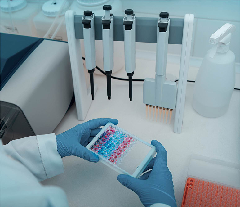 Figure 1. Determination of Protein Concentration by the Lowry Method.
1. Spectrophotometer absorbing light in the visible range and 1 mL plastic or glass cuvettes.
2. Copper reagent: Dissolve 20g of Na2CO3 in 260 mL water, 0.4 g CuSO4 · 5H2O in 20 mL water, and 0.2 g sodium potassium tartrate in 20 mL water and mix.
3. SDS solution: 1.0 g sodium dodecyl sulfate in 100 mL water (1 % w/v).
4. NaOH solution: Dissolve 4 g NaOH in 100 mL water to make a 1 M solution.
5. 2× Lowry working reagent: Mix three parts copper reagent with one part SDS solution and one part NaOH solution. This reagent is stable for 3 weeks.
6. 0.2 N Folin reagent: Mix 10 mL Folin reagent with 90 mL water. The reagent is stable for several months if stored in an amber bottle.
1. Set the spectrophotometer to read at 750 nm allowing 15 min for the instrument to equilibrate.
2. Prepare a bovine serum albumin (BSA) protein standard at 1 mg/mL concentration in duplicate. Dilute the protein standard in a volume of 0.4 mL to give fi ve concentrations over the range of 10–100 μg protein. Perform the dilution and readings for both standard solutions to give four readings at each protein concentration.
3. To 0.4 mL protein sample add 0.4 mL of the Lowry working reagent and incubate for 10 min at room temperature.
4. Add 0.2 mL of the 0.2 N Folin reagent, vortex, leave for 30 min, and read the absorbance values at 750 nm in the spectrophotometer. Readings can be done between 650 and 750 nm, but are more sensitive at 750 nm.
1. Advantages of the Lowry method: (a) The method is 100-fold more sensitive than the original biuret reagent, and (b) the method is more sensitive than determining absorbance at 280 nm.
2. Disadvantages of the Lowry method: (a) Many common substances (K+ , Mg2+, NH4+ , EDTA, Tris, carbohydrates, and reducing agents) interfere with the method; (b) the Folin reagent is reactive for only a short period of time after addition; (c) the method is complicated and requires more steps and reagents than the BCA or Bradford assays, and (d) the method is destructive to proteins; that is, once the protein sample has reacted with the dye, the protein cannot be used for other assays.
3. It is advisable to dissolve the protein standard in duplicate. If a protein standard is weighed or solubilized incorrectly in a single preparation, then all protein determinations will be inaccurate.
4. Protein determinations with the Lowry method should always be considered as a relative concentration of protein. It is accepted practice to express the relative concentrations of proteins during protein purification steps, provided that all protein concentrations are determined with the same method.
5. The Lowry determination methods produce a nonlinear curve, whilst absorbance at 280 nm produces a straight line as protein concentration increases.
6. The Lowry protein curves all proceed from the origin of the graph, provided that the correct "blank" or "control" has been used.
Reference
Loading ......
