
Inquiry Basket
 Loading ......
Myeloid-derived suppressor cells (MDSC) have been a target for basic and clinical studies related to chronic inflammatory diseases such as autoimmunity, chronic obstructive pulmonary disease (COPD), transplantation, infections, and cancer. In cancer, MDSC play an important role in limiting immune responses and their accumulation is associated with tumor growth, deficient response to immunotherapy, and poor survival. Flow cytometry is a powerful tool for detailed analysis of MDSC. Although surface markers have been described and extensively utilized for the identification of MDSC in human samples as well as in mouse models of research, the strategy for isolating and analyzing those cells by flow cytometry differs between research groups.
The precise identification and phenotypification of MDSC are crucial for understanding their role in the pathophysiology and predict the outcome of the disease. MDSC are divided into three major subtypes based on their cell surface phenotype: granulocytic polymorphonuclear MDSC (PMN-MDSC), monocytic MDSC (M-MDSC), and early-stage MDSC (e-MDSC). Human PMN-MDSC are defined as CD11bdim(or alternatively CD33dim), CD14neg, CD66bpos(or CD15pos), HLA-DRneg whereas M-MDSC are CD11bhigh, CD14pos, HLA-DRneg/Low, CD66bneg. Early-stage MDSC (e-MDSC), a less characterized subtype is CD11bdim, CD14neg, CD66bneg, HLA-DRneg. Mouse MDSC are identified by expressing CD11b and Gr1, which is comprised of two components, Ly6C and Ly6G. The mouse PMN-MDSC subset is identified as CD11bpos, Gr1pos, Ly6Cdim, Ly6Gpos, while M-MDSC are CD11bpos, Gr1pos, Ly6Chigh, Ly6Gneg. The minimum criteria for defining MDSC include these phenotypic surface markers; however, to fully define these cells as MDSC, assessing the functional and, if possible, biochemical and molecular features should be incorporated. Despite the cost, the long period of coculture, and thereafter the evaluation of T cell response, functional analysis of the immunoregulatory activity of MDSC on T cell proliferation is frequently performed in research with rodents. Unfortunately, it is less common with human samples for practical reasons, for example, most likely insufficient amount of samples to purify MDSC and T cells.
MDSC are functionally suppressor cells by producing antiinflammatory cytokines, arginase-1 and reactive oxygen species (ROS) such as superoxide. Also, MDSC express inducible nitric oxide synthase (iNOS) and programmed death-ligand 1 (PD-L1), among other molecules.
Arginase-1 is an enzyme that converts L-arginine into L-ornithine and urea, leading to the depletion of L-arginine. The absence of this amino acid impairs the proliferation and function of T and NK cells abrogating the protective immune responses. ROS critically affects the functions of T cells, their proliferation, differentiation, and survival.
Here we present a protocol to identify and quantify MDSC from peripheral blood and dissociated tissue (e.g., tumor) from human and mouse by using a panel of antibodies that collects the minimal recommended surface markers to characterize the phenotypic features of MDSC. This panel includes fluorescently conjugated antibodies against CD3, CD19, CD20, CD56 (lineage cocktail), CD45, CD33, HLA-DR, CD66b, and CD14 in combination with anti-arginase-1 or with the fluorescent probe dihydroethidium (DHE) to indicate cellular superoxide content. With this immunophenotypic analysis, the presence of MDSC with potential suppressive capacity can be evaluated by flow cytometry. Also, this approach can be used as the basis for developing additional panels where can be included other relevant molecules such as PD-L1, LOX-1, iNOS, as well as additional ROS indicators such as mitochondrial O2-(Mitosox) or intracellular H2O2 (DCFDA).
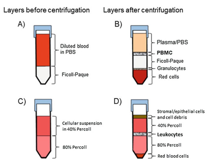 Figure 1. Schematic illustration of isolating of PBMC from whole peripheral blood by a density gradient centrifugation using Ficoll-Paque and tissue/tumor infiltrating-leukocytes by discontinuous Percoll gradient.
The detection of intracellular proteins such as arginase-1 (see Note 15) by flow cytometry requires cell fixation and permeabilization (see Note 16).
Acquisition of Data in the Flow Cytometer and Analysis
Reference
Loading ......
