
Inquiry Basket
 Loading ......
Far-western blotting is a method of characterizing protein–protein interactions, in which protein samples of interest are separated by gel electrophoresis, immobilized on a membrane, and then probed with a non-antibody protein. The term "far-western" was derived from western blotting, a similar method in which mem branes are probed directly with specific antibodies, and is also referred to as a west-western or blot overlay assay. Non-antibody proteins have been also used as a means to screen phage based expression libraries. Far-western blotting is very different from other commonly used methods to detect and characterize protein–protein interactions, and therefore complements these other approaches. Because the probe protein directly binds to denatured/separated proteins immobilized on a membrane, far-western blotting detects only direct interactions; by contrast, most non-far-western protein binding assays, such as immunoprecipitation and pull-down assays, may detect both direct association (two proteins make contact directly) and indirect association (two proteins do not make con tact, and another molecule in the ternary complex mediates the association). Thus, the far-western assay is often used to confirm direct interaction following immunoprecipitation or pull-down assays. The ability of far-western blotting to detect direct interactions is offset by limitations in the types of protein–protein interactions that can be detected. Because target proteins in a cell lysate are usually denatured in the process of gel electrophoresis, it may be difficult or impossible to detect interactions that require the native, folded conformation of the target protein. For this reason, far western blotting has been particularly useful in characterizing the binding partners of modular protein binding domains that bind to short, linear peptide motifs. It is now apparent that many signaling proteins interact with their partners via such modular binding domain–peptide interactions, and thus, the far-western approach is quite useful for analysis of signaling networks. However, these differences highlight the importance of using multiple approaches to assess specific protein–protein interactions. In far-western blotting, either a whole protein or fragment of a protein containing a suspected binding interface is used to probe interaction partners immobilized on a membrane. The interaction is visualized by direct labeling of the probe or by its subsequent detection with antibodies. There are a number of considerations in selecting the specific probe. First, ease of growth and purification of the probe must be considered. For the sake of cost and convenience, expressing probe proteins in bacteria is advantageous. However, only relatively small proteins (less than ~100 kDa) tend to remain soluble when grown in bacteria, so in general a fragment of a protein containing only the known or suspected binding domain will be easier to work with than the full-length protein. Second, it is useful to fuse the probe protein or domain to a tag sequence for ease of purification and detection. We routinely make probe proteins as glutathione S-transferase (GST) fusions, which has the dual advantage of allowing purification of proteins on glutathione columns, and allowing detection of bound probe with glutathione conjugates or with anti-GST antibodies. A further advantage of GST fusions is that GST exists as a stable dimer in solution. As in the case of antibodies, a dimeric probe binds with much greater avidity compared to a monomer to targets containing multiple binding sites, such as a membrane surface bearing many molecules of a target protein. In contrast to western blotting where a target protein is usually known in advance, far-western blotting can detect proteins on the basis of presence or absence of binding sites without any previous knowledge about their identities. From the intensity of bands observed on a far-western blot of a complex mixture of proteins, one can gain insight into both the number and relative affinity of binding partners for the probe in that sample. Furthermore, since some protein binding domains recognize their targets only after specific post-transcriptional modifications, far-western blot ting can be used to assess the modification status of multiple proteins in a sample. In this chapter, we will present a specific example of the utility of far-western blotting methods, in which GST-tagged Src homology 2 (SH2) domains, which bind specifically to tyrosine-phosphorylated target proteins, are used to probe the state of tyrosine phosphorylation of cellular proteins. Since quantitative comparison of interactions between multiple SH2s or other signaling proteins and their ligands is often of interest, we also provide a batch quantification method for multiple far-western blots.
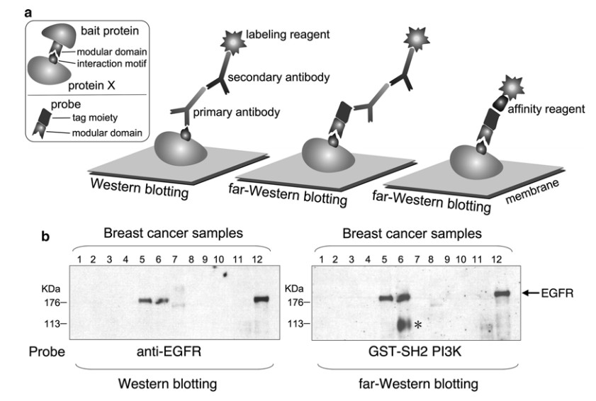 Figure 1. Comparison of western and far-western blotting.
Same as Subheadings Far-Western Blotting and Stripping and Reprobing.
As for western blotting, protein samples are separated by SDS- PAGE and transferred to a nitrocellulose or polyvinylidene fluoride (PVDF) membrane. The blocked membrane is then incubated with a probe followed by appropriate wash, and bound probes are visualized. Generally any protein samples compatible with western blotting can be used including whole cell lysates, purified proteins, and native or denatured samples. The far-western method described here is a rapid and simple protocol in which the membrane is prepared without denature–renature procedures, the probe is labeled directly, and probing is performed in one step. This protocol has been optimized to detect in vitro interaction between modular binding domain probes and immobilized proteins containing short peptide motifs (see Note 1). Of note, alternative protocols are available including another example in which proteins containing modular domains on a membrane are probed with labeled binding motifs. Below, we present a specific protocol for generating GST-SH2 domain probes and using them to probe tyrosine phosphorylated whole cell lysates. We have also included a method for aligning and quantifying multiple far-western blots which allows for comparative assessment of SH2 binding. Of course these procedures can be adapted for any modular protein binding domain and its binding partners with minor modifications. For all far-western blotting methods, detection of specific signal is strongly dependent on the quality of the probe protein. Insolubility, aggregation, or denaturation of the protein tends to cause nonspecific background, and even the native probe may bind nonspecifically to abundant proteins in the sample. Thus, it is important to: (1) confirm purified probe is soluble, folded, and not significantly degraded; (2) evaluate activity of the probe and optimize binding conditions if needed; and (3) always include appropriate positive and negative controls for each experiment to ensure any positive signal is indeed specific. To address these considerations, in the following section we will describe a detailed protocol for generation and evaluation of GST-SH2 fusion probes. Appropriate controls should be prepared considering the intended physiological activity of the probe. At minimum, GST alone, or more ideally GST fused to the domain of interest bearing a mutation known or suspected to abolish specific binding activity, should be used as a negative control probe.
Protein expression and solubility of GST-SH2 clones can be tested quickly in small-scale bacterial cultures (see Note 7), and the activity of the probe can be tested at the same time by a control pulldown assay.
Steps 16-19: Evaluate pTyr binding activity by GST pull-down assay (optional).
GST-SH2 probe is purified following the standard protocol for preparation of GST fusion proteins using pGEX series bacterial expression vectors (see Note 13).
Generally, fresh membranes are best for far-western blotting; stripping and reprobing of the membrane may result in significant signal loss and increased non-specific background. Nevertheless recycling membranes might be beneficial if sample is limited, or if precise comparison of specific bands is needed within the same membrane.
Although a single far-western blot is useful for identifying differences between samples, comparison of probe specificities, such as the phosphosite preference of different SH2 domains, requires parallel probing and quantification of multiple replica membranes. Here we describe a method for the preparation, probing and detection, image adjustment, and batch quantification of multiple membranes.
For accurate batch quantification, all images must be well aligned. Images from the same physical membrane, e.g., reprobing, can be easily aligned using the frame position marks, while those from different membranes must be carefully aligned using the anti-phosphotyrosine or other reference blot images.
Bin-based batch quantification partitions each far-western lane into a set number of equal sized rectangular regions of interest using a grid. This method allows for an unbiased quantification of far-western blots by capturing the differences in SH2 binding patterns produced by each probe and compensating for minor residual variations in band position. Since all blots are quantified as a batch, this method also ensures every far-western is partitioned equally and quantified using the same parameters. While there are multiple possible ways to partition far- western data for quantification (i.e., molecular weight standard or band-based) we have found that a bin-based quantification works well when gels/blots are relatively homogenous in shape and have many bands that need to be quantified.
Addgene.
NCBI Entrez Gene.
ScanProsite.
The Human Protein Reference Database.
UniGene.
SH2 domain information site.
Reference
Loading ......
