
Inquiry Basket
 Loading ......
Dendritic cells (also known as accessory cells) are highly specialized antigen-processing and antigen-presenting cells that are located in mucosal tissues, skin, and lymphoid tissues. Dendritic cells play a major role in the initiation of immune responses because they act as messengers between the innate and adaptive immune systems. The main function of DC cells is to process and present antigens on their cell surface leading to activation of T cells. Dendritic cells-based immunotherapy is a cutting-edge approach to disease treatment that harnesses the power of the patient's own immune system.
Dendritic cells (DCs) were derived from pluripotent hematopoietic stem cells (HSCs) of the bone marrow. They belong to a group of antigen-presenting cells (APCs) that include B-cells and macrophages. They were originally discovered in 1973 by two Canadian scientists named Ralph Steinman and Zanvil Cohn as a previously undefined cell type in the mouse spleen, and eventually known as "dendritic cells" because of their typical features of multiple extended dendritic or pseudopodia-like cytoplasmic protrusions during their maturation stage.
DCs are found in tissue that has contact with the outside environment, such as lung mucosa, epithelial cells of the skin, and the linings of the nose and the gastrointestinal tract. These regions form the interface between the body and the environment and are constantly exposed to foreign proteins and pathogens. Immature forms of DCs are also found in the blood. Once activated DCs move to the lymph tissue to interact with T and B cells and help shape the adaptive immune response. Therefore, DCs are considered to be the center of the immune system, because they provide an important link between the innate and adaptive immune response.
DCs are a heterogeneous group of cells consisting of subsets. They can be broadly classified as conventional DCs consisting of two subsets (DC1 and DC2), plasmacytoid DCs, inflammatory DCs, and Langerhans cells.
Conventional DCs (Classical DCs, cDCs)
cDCs can be divided into two major subsets, based on the expression of the following surface molecules. These include CD8α+ and CD103+ or CD11b. In addition, both subsets can be found both in the lymphoid tissues, such as the spleen, lymph nodes, and bone marrow, and in most of the non-lymphoid tissues.
The CD8α+ and CD103+ cDCs are the best-characterized cDC subsets so far, both in terms of phenotype and gene expression signature. Throughout evolution, they appear to be conserved. Functionally, the splenic CD8 cDCs are specialized in cross-presentations of exogenous antigens on MHC-I molecules to CD8 T-cells. Additionally, the skin-derived CD103 cDCs have been described to display cross-presentation activity.
The most abundant cDCs in lymphoid organs are the CD11b+ cDCs, except in the thymus, and they have also been found in non-lymphoid tissues. Currently, CD11b+ is defined as heterogeneous and remains less well characterized compared to CD8α+ and CD103+ DCs. Monocyte-Derived DCs (Inflammatory DCs, moDCs)
Monocyte-derived DCs (moDCs) are also known as "inflammatory DCs". They arise from the myeloid progenitor and play a vital role in the immune response, because during the onset of infection they tend to provide a pool of APCs that can effectively initiate an adaptive immune response. During inflammation or infection, blood circulating monocytes that are normally known to express receptors for Granulocyte Macrophage-Colony Stimulating Factor (GM-CSF), Macrophage-Colony Stimulating Factor (M-CSF), and Interleukin-4 (IL-4), other differentiation, and chemoattractant molecules tend to invade tissues and are attracted to the site of inflammation, where they are subsequently differentiated into moDCs. However, during an infection, upon entry of moDCs to the site of infection, the moDCs produce TNF-α and an inducible nitric oxide synthase (iNOS), which eventually confers innate protection.
Plasmacytoid Dendritic Cells (pDCs)
pDCs arise from the lymphoid progenitor. They are found throughout the body, circulating in the blood and peripheral organs, and reside within the lymph nodes, spleen, thymus, and bone marrow (BM). The pDCs display a distinct morphology and surface phenotype, and possess a highly developed secretory compartment. They are characterized by their round shape and plasmacytoid morphology, and have a resemblance to lymphocytes, while the IL3/CD40L cultured pDCs have a microscopic appearance that is similar to that of moDCs. pDCs are generally dormant, but upon stimulation by toll-like receptors (TLR7 and TLR9), they secrete copious amounts of interferon-gamma, which plays a major role in viral and bacterial infections.
Langerhans or Epidermal DCs
Langerhans cells are a distinct population of mononuclear phagocytic cells that reside within the epidermis. They were the first DC subset to be discovered, in the 19th century, and are considered to be primary sentinel tissue-resident DCs that sample their environment. Upon activation, Langerhans cells (LCs) tend to migrate via the dermis into the nearest regional lymph nodes and eventually present antigens to naïve T-cells. Therefore, this type of DC subset is categorized as "migratory DCs" and other non-lymphoid-tissue-resident DCs, such as dermal and intestinal DCs, also belong to this category of DC subsets.
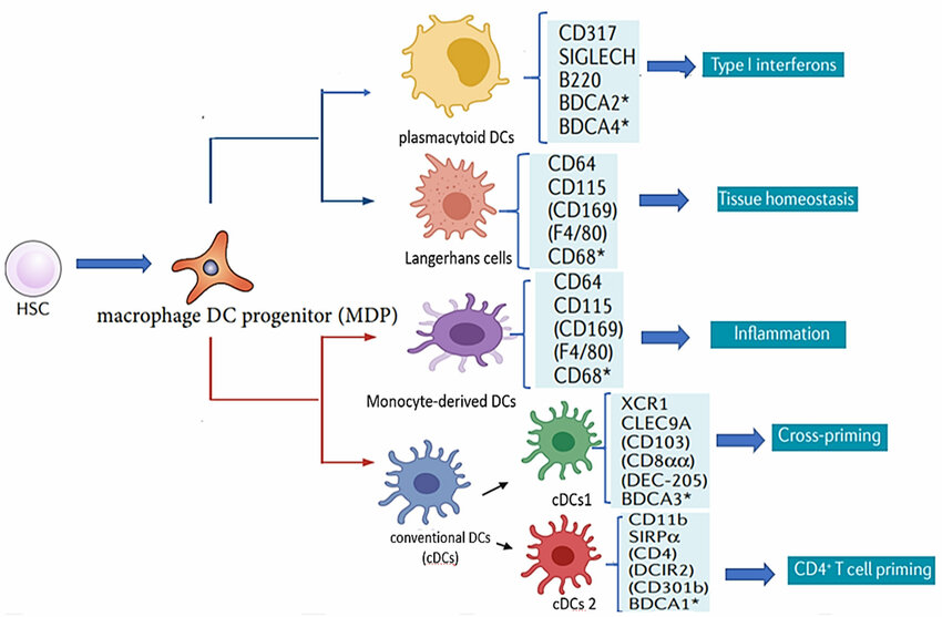 Fig. 1 Dendritic cell (DC) subsets and their corresponding functions
To date, no single cell marker has been found to be expressed exclusively on DCs, therefore a combination of the presence and absence of various cell markers can be used to identify the DCs. A combination in expression of CD1c, CD11c, CD16, CD45, CD141, CD370 (CLEC9A), HLA-DR, and lineage exclusion markers CD3, CD14, CD19, CD20, CD56 can be used to differentiate between the main subsets and other types of cells.
Dendritic Cell Therapy (DCT) is a cutting-edge immunotherapeutic approach that leverages the unique properties of dendritic cells (DCs), the most potent professional antigen-presenting cells in the immune system, to treat various diseases, particularly cancer. DC therapy has been utilised in medicine since 2010. Over 4,500 humans have passed through it since then. It is efficaciously used in the remedy of breast cancer, prostate cancer, sarcoma, melanoma, glioma and different cancers.
DC-based immunotherapy is a cutting-edge approach to disease treatment that harnesses the power of the patient's own immune system. The process involves isolating DCs from the patient's blood or tissue and then modifying them in the laboratory to present tumor-specific Ags.
DCs have shown therapeutic effects in cancers, such as colorectal, pancreatic, and breast cancer, although DC-based immunotherapy has not achieved the expected efficacy in most solid tumors. Coadministration of cytokines with DC vaccination has shown promising results, significantly suppressing tumor growth and improving in vivo immune responses. To enhance the longevity of DCs in vivo and promote increased migration, one approach is to coadminister cytokines, such as inflammatory cytokines (e.g., interleukin (IL)-2, IL-12, and IFN), as adjuvants with a DC vaccination. Thus, DC vaccination that enhances DC migration through coadministration with cytokines may become a novel approach in cancer immunotherapy.
DC-Based Treatment of Inflammatory and Autoimmune Diseases
Conventional treatments for autoimmune diseases involve suppressing general immune function to modulate uncontrolled inflammation. Tolerogenic DCs (tDCs) are characterized by reduced expression of costimulatory molecules and secretion of proinflammatory cytokines, such as IL-12, while exhibiting elevated production of anti-inflammatory cytokines, such as IL-10. For example, CD103+ DC, one of the cDC2 subsets in the gut, can promote immune tolerance by inducing regulatory T (Treg) cells. In the mouse spleen, the cholesterol regulatory pathway that involves liver X receptor (LXR) α/β also can control the immunogenic and tolerogenic cDC maturation. These tDCs are instrumental in maintaining immune homeostasis by activating Treg cells, inhibiting effector T cells, and modulating Th1/Th2 immune responses. tDCs play an important role because they are used as anti-inflammatory and immunosuppressive targets in several models of autoimmune diseases, contributing to the induction of peripheral tolerance. tDC-based therapy is applicable to autoimmune diseases, including rheumatoid arthritis (RA), systemic lupus erythematosus, Crohn's disease, and type 1 diabetes mellitus (T1DM).
Loading ......
