
Inquiry Basket
 Loading ......
The discovery of embryonic and adult stem cells has led to great interest and excitement about the therapeutic potential of these cells across medical disciplines. The two hallmark properties of stem cells are their ability to undergo self-renewal and their potentiality for differentiation into the multiple cell lineages specific from the organ from which they derive. Because of these properties, stem cells are essential for an organism to maintain tissue homeostasis and for tissue repair following injury. The features of self-renewal and pluripotentiality also make adult stem cells invaluable therapeutic tools for reversing the symptoms/pathology of neurodegenerative disease, promoting tissue repair after ischemia or stroke, or as a model system to understand how to inhibit the proliferative capacity of recently identified tumor-associated stem cells.
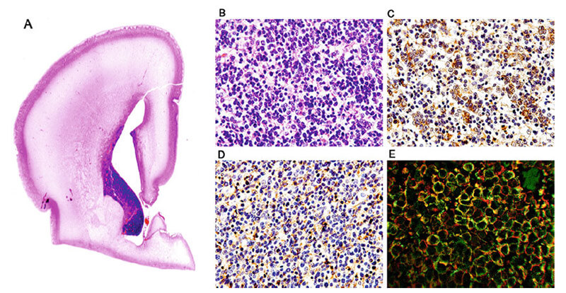 Figure 1. Germinal Matrix.
These cells may be isolated and cultured in vitro so that they can be manipulated and studied by researchers. Neural stem cells are present in two specialized niches in the adult mammalian brain, the dentate gyrus of the hippocampus and the germinal matrix, located in subventricular zone of the lateral ventricles. Once removed, they can be propagated as adherent cells in traditional two-dimensional cultures or embedded in extracellular matrices, or as floating spheres called neurospheres. Culturing neural stem cells in the form of neurospheres helps to select for and preserve stem cell-like properties of isolated cells, and neural stem cells grown as neurospheres can be maintained in this primitive state for many passages, creating a readily available, stable pool of stem cells for research. The cells can be used to ask defined questions about factors that alter stem cell properties, such as their capacity for self-renewal, or how experimental or genetic manipulations affect the neuronal or glial differentiation program.
In order for these types of experiments to be successful, researchers must be able to definitively identify the genetic and phenotypic changes resulting from the experimental manipulations. Immunocytochemistry is an invaluable tool for cell phenotyping. Neural stem cells and their progeny can be identified by the use of select protein biomarkers. These biomarkers are selectively expressed by stem, neuronal, or glial cells; or during specific stages in the differentiation process.
Neurosphere Growth Medium: neurobasal medium supplemented with 20 ng/ml EGF, 20 ng/ml bFGF, 2 mM GlutaMAX, pen/strep, and 2% StemPro Neural Supplement.
Neurosphere Differentiation Medium: neurobasal medium supplemented with 10% fetal calf serum, 2 mM GlutaMAX, pen/strep.
Neuronal Differentiation Medium: neurobasal medium supplemented with 2% B27, 2 mM GlutaMAX, and pen/strep.
Oligodendrocyte Differentiation Medium: neurobasal medium supplemented with 2% B27, 30 ng/ml T3, 2 mM GlutaMAX, and pen/strep.
Astrocyte Differentiation Medium: neurobasal medium supplemented with 10% FCS supplement, 100 ng/ml CNTF, 2 mM GlutaMAX, pen/strep.
When the neurospheres are ready to be passaged, gently suspend the neurospheres, and transfer the media and suspended neurospheres into a sterile conical tube.
Neurospheres from passages 1–3 (P1-P3) are often irregularly shaped and disorganized clusters, with debris still present from the isolation procedure. After P3, healthy neurosphere should be shiny, smooth with a uniform outline when viewed through a tissue culture microscope. When Neurospheres reach this state, they are ready for downstream studies.
The plating density of the neural stem cells in later passages is highly dependent on the question of the experiment. If the experimental outcome is to determine the clonality of isolated or treated cells, the cells must be plated at very low densities to endure that the resulting neurospheres are the result of single cell expansion and not the result to aggregation of multiple cells and smaller neurospheres. The most stringent plating method calls for cells to be plated in 96-well plates at very low plating densities (1–10 cells/μl with 200μl/well). The researcher can then identify which wells contain only a single cell and monitor just those wells for neurosphere formation. If the experiment is designed to measure other outcomes such as the influence of growth factors or pharmacologic treatments on neuronal development, then less stringent plating conditions are required (see Note 5).
Coat Chamber Slides or Coverslips with Poly-L-Ornithine/Laminin
Differentiation of Intact Neurospheres
The neurospheres are ready for plating when they reach approximately 150 nm in diameter (between day 6–8 of culture).
Differentiation of Dissociated Neurospheres
When neurospheres are ready for harvesting, precoat glass coverslips or chamber slides and collect neurospheres as described above.
The resulted cell population will reflect the potentiality of the stem cell culture and can be used to test the effects of genetic or therapeutic manipulation on neural stem cell fate. Alternatively, culture conditions can be adjusted to selectively push neural stem cells into different lineages, as outlined below.
Culture Conditions to Preferentially Differentiate Neural Stem Cells into Neurons
Culture Conditions to Preferentially Differentiate Neural Stem Cells into Astrocytes
Culture Conditions to Preferentially Differentiate Neural Stem Cells into Oligodendrocytes
Fixation with Paraformaldehyde
Fixation with Methanol
Fixation with Acetone
The choice of fixation method used will depend on both the epitope and primary antibody and therefore this step may require some optimization to determine which method is best. Fixation using cross-linking reagents, such as paraformaldehyde are better at preserving cell structure, but may reduce the detection of some targets as cross-linking may interfere with antibody binding. Organic solvents like acetone or ethanol dehydrate and precipitate proteins on the cellular architecture and tend to work well for detection of cytoskeletal components but may reduce detection of soluble proteins.
The neurospheres can be fixed and immunolabeled as intact neurospheres. This may be desired if the goal is to identify pathways associated with maintenance of, or the differentiation from, the stem cell phenotype. Immunocytochemical analysis of the intact neurosphere would allow the investigation of how the expression of proteins of interest changes during the differentiation program, or to identify the presence of representative markers of apoptosis or cell cycle regulation following experimental manipulation of the neurosphere cultures. Identifying the patterns of protein expression in intact neurospheres is advantageous since dissociation and plating of the neural stem cells initiates differentiation and alters gene expression. In addition, a model containing the majority of cells present in the brain allows one to track cytopathological changes to specific phenotypes, and to observe the interactions between different cell types.
Fixation and Permeabilization of Intact Neurospheres
Neurospheres Generated from Brain Tumor Stem Cells Neurosphere assays can be utilized to identify neural stem-like properties in cells derived from primary brain tumors. Primary tumor cells have the propensity to retain their morphological and physiological feature when maintained in neurosphere culture as compared to cells cultured in two dimensional cell cultures. Like neural stem cells, neurosphere-forming cells derived from primary tumors are self-renewing and retain the ability to differentiate into the cells found in the tumor in vivo. In addition, upon transplantation into nude mice, they form tumors that reflect the tumor cell composition from which they were derived. The propensity of primary tumor-derived cells to form neurospheres has been correlated with clinical outcome in both pediatric tumors and glioblastomas. The outcome of genetic manipulation and/or pharmacological treatments on BTSC could be analyzed using neurosphere assay. Immunocytochemical analysis of BTCS-derived neurosphere could identify whether tumor stem cell markers are altered or preserved.
Reference
Loading ......
