
Inquiry Basket
 Loading ......
The comet assay, also called the single-cell gel electrophoresis, is an invaluable tool for quantifying DNA damage in individual cells. The concept of DNA migrating in an agarose gel by an electric current was first introduced in 1984 by Ostling and Johanson as a method to measure single stranded breaks that caused a relaxation of the DNA supercoiled structure under neutral conditions. They also introduced the terms "comet" and "tail" as they described the shape of the displaced DNA and the tail moment (tail length and percentage of fluorescence) as an indicator of DNA migration. A modified version to quantify low levels of DNA damage in single cells after exposure to X-rays and hydrogen peroxide under alkaline conditions was published by Singh et al. in 1988. The assay relies on the principle that low-molecular-weight DNA fragments migrate away from damaged supercoiled DNA under an electric current, creating in a visible DNA fragment tail off of the intact nuclear DNA head. Individual comets can then be scored qualitatively using manual analysis or quantitatively by using software that determines the relative fraction of DNA in the comet tail. The comet assay protocol is relatively simple and can be completed in 1 day with as few as 1000 cells, making it useful for monitoring the efficacy of genotoxic agents, such as chemotherapeutics and radiation, and for determining the rate of DNA repair in cancer cells. Modifications to the comet assay have enabled the detection of DNA damage associated with specific cellular processes and genes. For instance, the use of bromodeoxyuridine in combination with the comet assay facilitates the detection of DNA breaks in replicating cells, and by employing lesion-specific enzymes sensitivity and specificity of the comet assay can be increased. In addition, in situ hybridization can be also combined with the comet assay to identify DNA breaks in individual genetic regions.
Despite its utility, there are several limitations the comet assay, which need to be discussed. Small variations in agarose concentration, voltage gradients, current, and electrophoresis time can drastically affect experimental results. Care must be taken to minimize DNA damage during the protocol and samples must be viable, as necrotic or apoptotic cells cannot be analyzed. While there has been some success in using cryopreserved cells for the comet assay, it is suggested that live cells are prepared immediately before use to ensure experimental quality and reproducibility. The number of cells able to be analyzed in 1 day is limited to 500–600 individual cells if performed by hand or 50–60 slides if using an automated software, reducing the utility of the comet assay for high throughput studies.
The comet assay may be performed under alkaline or neutral conditions. The neutral comet assay exhibits reduced sensitivity compared to the alkaline comet assay, but it detects solely double-stranded breaks, whereas the alkaline comet assay detects both single- and double-stranded DNA breaks. The principle of alkaline methods is that only if the DNA-base pairing is disrupted by an alkaline environment, the breaks can be detected. At neutral pH, however, the discontinuities in the single stranded breaks cannot be detected as the DNA is supercoiled and tightly packed, and in this condition only double stranded brakes will contribute to the formation of the comet. The methods for both alkaline and neutral comet assays are described herein.
Sample Preparation and Comet Assay
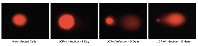 Figure 1. An example of single cell electrophoresis in glial cells exposed to JCPyV infection.
Reference
Loading ......
