
Inquiry Basket
 Loading ......
Campylobacter is a genus of Gram-negative bacteria with cells that are curved or spiral-shaped, typically exhibiting an S-shape or helical form. Under old culture conditions or stress, they may also transform into coccoid forms. Campylobacter are Gram-negative bacteria. They are rapidly motile with a polar flagellum, moving in a spiral manner. They are usually microaerophilic, but some strains are facultatively anaerobic or even aerobic. They are non-spore-forming. Campylobacter species are found in many warm-blooded animals and birds, with a particular prevalence in poultry. They are also an important zoonotic pathogen.
At present, 32 formally described species and 9 subspecies are recognized, distributed in five phylogenetic groups. The most important human pathogens of this genus are C. jejuni and C. coli. C. jejuni is the most frequent bacterial cause of gastroenteritis in humans. The frequency of isolation of this organism far exceeds the more familiar intestinal pathogens E. coli, Shigella and Salmonella. C. coli, the species most closely related to C. jejuni, causes 1% to 25% of all Campylobacter diarrhea. C. coli is closely related to C. jejuni and accounts for 1% to 25% of all Campylobacter associated diarrhea cases. In addition to C. jejuni and C. coli, 13 of the other 30 species have been rarely associated with human or animal infections as pathogens. Pathogenic C. fetus, C. upsaliensis, C. insulaenigrae, and C. cryaerophila, for example, have been reported to cause bloodstream infections, periodontal infections, abscesses, meningitis, diarrhea and gastroenteritis. The lack of genomic data for most of these species has made it impossible to extensively study the patterns of intraspecies genetic variation and genome evolution in them. The question of how many of these emerging Campylobacter species cause disease and how it spreads among the hosts is still unknown.
Table 1. Currently described Campylobacter species
| Campylobacter Species | Known Sources | Human Disease Associated |
| C. jejuni subsp jejuni | Poultry, cattle, sheep, wild birds, pigs | Gastroenteritis, meningitis, septicemia, Guillain-Barre syndrome |
| C. jejuni subsp doylei | Humans | Gastroenteritis, septicemia |
| C. coli | Pigs, poultry, sheep, wild birds, cattle | Gastroenteritis, septicemia, meningitis |
| C. lari subsp lari | Wild birds, poultry, dogs, cats | Gastroenteritis, septicemia |
| C. upsaliensis | Dogs, cats | Gastroenteritis, septicemia |
| C. lanienae | Cattle, pigs | Gastroenteritis |
| C. curvus | Humans | Periodontal disease, gastroenteritis |
| C. mucosalis | Pigs | None at present |
| C. avium | Poultry | None at present |
(Source: Fitzgerald C. 2015)
C. jejuni exhibits extensive genetic variation arising from both internal genomic mechanisms and gene exchange between different strains. Genome sequencing of C. jejuni has revealed numerous hypervariable sequences, mainly composed of homonucleotide repeats. Many of these hypervariable loci are found in genes for production or modification of the carbohydrate structures exposed on the cell surface such as capsules, lipooligosaccharides and flagella. C. jejuni is naturally competent for transformation, and thus can take up exogenous DNA from the environment. Recombination between strains has also been observed. Coupled with the frequency of transformation events, this could be responsible for the high level of genetic diversity that has been observed for this species. The frequency of natural transformation was also dependent on the concentration of carbon dioxide, as well as the bacterial cell density, which suggests that horizontal gene transfer by this mechanism may be environmentally regulated in vivo.
The lipooligosaccharides (LOS) of C. jejuni are highly diverse, which is closely related to their function in evading the immune system. Many C. jejuni LOS structures resemble neuronal gangliosides found in the human nervous system, and this molecular mimicry is considered one of the causes of autoimmune diseases. Mutations in LOS biosynthesis genes also have been shown to have an impact on the resistance of the organism to serum, and its ability to adhere and invade and human intestinal epithelial cell lines. The capsule of C. jejuni has also been shown to be important in serum resistance, epithelial cell adhesion and invasion, colonization of chickens, and virulence in a ferret model. C. jejuni strains have been serotyped for many years almost exclusively on the antigenic differences in capsule structure. So it is clear that the capsular polysaccharides are immunogenic and the extreme variability is likely an immune evasion strategy. Capsules may also be involved in protection against desiccation or bacteriophage infection, although this has not been well studied.
C. jejuni has two systems for protein glycosylation. O-linked glycosylation is the addition of glycans to serine/threonine residues of flagellar proteins, while N-linked glycosylation is the presence of glycans on asparagine residues of proteins. Since flagella and motility have a demonstrated role in C. jejuni biology, loss of motility mutations caused by O-linked glycosylation are also loss of adhesion and invasion to host cells and virulence in ferret models. N-linked glycosylation is highly conserved in all studied C. jejuni strains and C. coli, suggesting it plays a more fundamental role in C. jejuni biology.
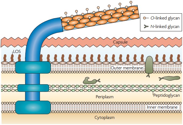 Figure 1. The C. jejuni glycome and surface structures
(Source: Young KT, et al. 2007)
Transmission of C. jejuni and C. coli is usually through the fecal-oral route. Ingestion of food or water contaminated with feces from an infected animal or human host or through direct contact with feces can all lead to infection. Consumption of undercooked poultry, untreated water and unpasteurized milk are food contamination methods. Campylobacteriosis is the most common reported bacterial foodborne illness. Epidemiologic data have demonstrated regional and population-based differences in the incidence of illness. Campylobacteriosis has shown seasonality with a sharp increase in cases during the warm months. A significant reason is the presence of ambient and water temperatures in the environment that are ideal for bacterial survival and transmission. Local differences in the incidence of campylobacteriosis have been observed, with some rural areas having a higher rate than urban communities, which has been ascribed to a greater exposure to animals, especially poultry and livestock and untreated water. Lockdown conditions during the COVID-19 pandemic reduced much of the human interpersonal contact and eating out, leading to a precipitous drop in the number of campylobacteriosis cases in 2020 worldwide, highlighting the role of these factors in its transmission.
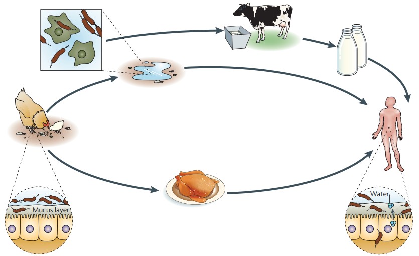 Figure 2. The sources and outcomes of C. jejuni infection
(Source: Young KT, et al. 2007)
Campylobacter infection leads to several diseases such as acute enteritis, extraintestinal infections, and post-infection sequelae. Campylobacter infection is self-limiting with 5 to 7 days of symptoms and most patients do not need antibiotic treatment, with complete recovery. The relapse rate is about 5% to 10% after the first illness. Symptoms occur 2 to 5 days after the ingestion of the bacteria. They are characterized by diarrhea with abdominal cramps and acute abdominal pain. Fever, nausea, and vomiting are common. Afebrile bloody diarrhea may occur in newborns and infants.
Occasionally, healthy individuals can acquire extraintestinal infections. On the other hand, it is very much more common in immunocompromised patients, elderly patients, and pregnant women. The most common form of extraintestinal infection is bacteremia, either diarrheal or non-diarrheal. Post-infectious sequelae of Campylobacter include GBS, reactive arthritis, and irritable bowel syndrome. C. jejuni is the most common bacterial cause of GBS, and 30% of those with GBS have a preceding history of infection with C. jejuni. Reactive arthritis occurs in 2% to 5% of cases, and about a third of patients develop irritable bowel syndrome.
C. jejuni can translocate from the apical surface of intestinal epithelial cells to the basolateral surface. When at the basolateral cell surface, C. jejuni can adhere to extracellular matrix protein fibronectin and invade epithelial cells. The route of translocation of C. jejuni remains a controversial issue. An early theory for this translocation process was a transcellular pathway, with the bacteria entering the host cell from the apical side of the cell, then traversing the cytoplasm to the basolateral side where they exit. A paracellular route of translocation has more recently been suggested, with the bacteria crossing between adjacent epithelial cells by passing through tight junctions and adherens junctions. C. jejuni was recently found to secrete the serine protease HtrA, which cleaves E-cadherin, and allowed for paracellular transverse of polarized MKN-28 cell monolayers. In a separate study, it was observed that during polarized human intestinal Caco-2 cell infection, HtrA cleaved the tight junction protein occludin, leading to a redistribution of occludin from the tight junction into the cytoplasm. C. jejuni invasion was enhanced in cells depleted of occludin, which suggests that C. jejuni-mediated tight junction disruption may facilitate penetration through to the basement membrane and invasion. Equivalent redistribution of occludin and tight junction associated protein ZO-1 was seen in biopsies taken from campylobacteriosis patients, confirming the damage of tight junctions during infection, and supporting the model of paracellular translocation. In addition, C. jejuni can also trigger non-invasive E. coli and Lactococcus lactis to translocate towards the basolateral region through an HtrA-dependent mechanism. This has raised the possibility that the HtrA of C. jejuni is a key factor for eliciting pathogen-associated abnormal immune responses in gut microbiota.
 Figure 3. The proposed translocation and invasion mechanisms utilised by C. jejuni
(Source: Tikhomirova A, et al. 2024)
C. jejuni is a facultative intracellular organism that can persist in intracellular compartments of intestinal epithelial cells. Specialized structures named Campylobacter-containing vacuoles enable this by avoiding fusion with lysosomes. Bacterial invasion of host cells typically occurs via two mechanisms: the "zipper" and the "trigger" methods. Zipper mechanism: Bacterial adhesins stick to host cell receptors. This causes rearrangement of cytoskeleton of the host cell and engulfment of the bacterium by endocytosis. Trigger mechanism: A type III secretion system injects effector proteins into the cytosol of the host cell. This activates signaling cascades which results in membrane ruffling and uptake of the bacteria. This process resembles macropinocytosis. C. jejuni's invasion mechanism combines features of both. Numerous surface molecules promote its adhesion to and invasion of intestinal epithelial cells. Adhesion helps the bacteria initially attach, preventing clearance by gastrointestinal motility and providing a platform for effector protein delivery. Some proteins, such as CadF, FlpA, JlpA, Peb1 and Peb3, are suspected adhesins. CadF and FlpA specifically bind extracellular matrix protein fibronectin on the basolateral surface of intestinal epithelial cells, resulting in the activation of phosphorylation of the focal adhesion protein paxillin and downstream signaling to regulate membrane trafficking and cytoskeletal rearrangement to promote invasion. In FlpA-deficient mutants, delivery of the effector protein CiaC is reduced, indicating that adhesion is critical for effector protein translocation.
References
| Target | Cat. No. | Product Name | Size | Species | Application | Detection Sample | |
| Campylobacter | DEIA-NS2401-17 | Campylobacter IgA ELISA Kit | 96T | Human | Qualitative | Serum, plasma | Inquiry |
| DEIA-NS2401-18 | Campylobacter IgG ELISA Kit | 96T | Human | Qualitative | Serum, plasma | Inquiry | |
| DEIA2560 | Campylobacter ELISA Kit (Fecal) | 96T | Human | Quantitative | Feces | Inquiry | |
| C. jejuni | DEIA-XY89 | Campylobacter jejuni IgA ELISA Kit | 96T | Human | Quantitative | Serum, EDTA plasma, heparin plasma, citrate plasma | Inquiry |
| DEIA-XY90 | Campylobacter jejuni IgG ELISA Kit | 96T | Human | Qualitative | Serum or plasma (EDTA, citrate, heparin). | Inquiry | |
| DEIA-XY91 | Campylobacter jejuni IgM ELISA Kit | 96T | Human | Quantitative | Serum, EDTA plasma, heparin plasma, citrate plasma | Inquiry |
| Target | Cat. No. | Product Name | Host | Isotype | Application | |
| C. jejuni | CABT-L3146 | Mouse Anti-Campylobacter jejuni / Campylobacter coli monoclonal antibody, clone D12057N | Mouse | IgG2b | LFIA | Inquiry |
| CABT-L3147 | Mouse Anti-Campylobacter jejuni / Campylobacter coli monoclonal antibody, clone D12058N | Mouse | IgG1 | LFIA | Inquiry | |
| Campylobacter | DPAB-CS23097 | Goat Anti-Campylobacter Species Polyclonal antibody | Goat | IgG | ELISA | Inquiry |
| CABT-CS110 | Mouse Anti-Campylobacter Porin Monoclonal antibody, clone DK17 | Mouse | IgG1 | WB, IB, IC | Inquiry | |
| DPAB31490 | Anti-Campylobacter Polyclonal antibody | Rabbit | IgG | ELISA, WB, IF | Inquiry | |
| CABT-RM188 | Magic 6 4 Mouse Anti-Campylobacter Monoclonal antibody, clone 39U6C0 | Mouse | IgG1 | ELISA (cap), LFIA | Inquiry | |
| CABT-RM189 | Magic 6 4 Mouse Anti-Campylobacter Monoclonal antibody, clone 39U6C1 | Mouse | IgG2b | ELISA (det), LFIA | Inquiry |
| Target | Cat. No. | Product Name | Expression System | Tag/Conjugate | Application | |
| C. jejuni | DAG-WT3805 | Recombinant Campylobacter jejuni CdtB protein [His] | E. coli | His | ELISA, WB | Inquiry |
| DAG-WT3676 | Inactivated Campylobacter jejuni Culture Fluid (AS-83-79) | N/A | N/A | Control | Inquiry | |
| DAG-WT3675 | Inactivated Campylobacter jejuni Culture Fluid (CIP 702) | N/A | N/A | Control | Inquiry | |
| DAGC033 | Recombinant Campylobacter jejuni (strain RM1221) Flagellar L-ring protein | E. coli | Unconjugated | N/A | Inquiry | |
| C. hominis | DAG-ZL0289 | Inactivated Campylobacter hominis Culture Fluid | N/A | N/A | Control | Inquiry |
Loading ......
