
Inquiry Basket
 Loading ......
Human chorionic gonadotropin (hCG) can be synthesized by cells such as chorionic syncytiotrophoblasts, a variety of primary non-trophoblastic tumors, the anterior pituitary gland, and cytotrophoblasts. Different cell types produce different subtypes of hCG, which have different biological functions. The main functions of hCG synthesized from the trophoblast cells of the chorionic complex include the promotion of progesterone production by the luteal cells and the subsequent growth of the cytotrophoblast cells. Highly glycosylated hCG in trophoblast cells promotes the growth and invasion of these cells, leading to choriocarcinoma cells, and hCG synthesized by nontrophoblastic tumors free β-subunit suggestive of malignant cancer and poor prognosis. The hCG synthesized by the anterior pituitary gland is low throughout the menstrual cycle and acts similarly to luteinizing hormone (LH).
The hCG hormone consists of a α subunit and a β subunit, which are linked by monovalent hydrophobic and ionic interactions. All isoforms contain the alpha subunit, which is also present in other hormones such as LH, follicle stimulating hormone (FSH) and thyroid stimulating hormone (TSH). The hCG hormone is distinguished from other hormones by the β-subunit. The different isoforms of hCG differ structurally, and these forms include: intact biologically active heterodimeric hCG (hCG), nicked hCG (hCGn), free hCG β-subunit, nicked free hCG β-subunit (hCGβn), core fragment of hCG β-subunit (hCGβcf), highly glycosylated hCG (hCG-H), and sulfated hCG.
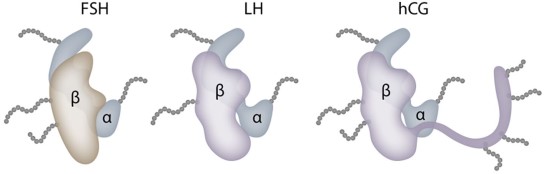 Figure 1. Gonadotropin molecules
(Source: Oyatogun O, et al. 2021)
hCG is a pregnancy-specific hormone that is essential for the development of the fetus and placenta. one of the most important functions of hCG is to promote the production of progesterone, which strengthens the endometrial wall during pregnancy and prevents menstrual bleeding. In addition, it coordinates the growth of the fetus and the uterus, signals the endometrium of impending implantation, and participates in the regulation of trophoblast differentiation as well as angiogenesis and vasculogenesis in the uterine wall. hCG stimulates the production of endocrine gland-derived vascular endothelial growth factor (EG-VEGF), which acts on cellular trophoblast. Through this action, the trophoblast cells can form an embolus that prevents maternal blood from flowing into the chorionic villous space during early pregnancy. hCG also inhibits immune-mediated mechanisms to prevent placental tissue rejection. hCG was found to up-regulate anti-macrophage inhibitory factors during pregnancy, thereby decreasing macrophage activity at the utero-placental interface.
Table 1. Some forms of clinically relevant hCG and their functions
| hCG Type | Production | Function |
| Intact, biologically active heterodimeric, normally glycosylated hCG | Syncytiotrophoblast hydatidiform moles | Pregnancy:
|
| Hyperglycosylated hCG | Extravillous cytotrophoblast cells |
|
| Free hCG β-subunit | Pregnancy: Implanted blastocysts and trophoblasts Malignancies: choriocarcinoma, nonseminomatous testicular tumors, bladder, cervical, pancreatic, lung, ovarian, endometrial cancers | Pregnancy:
|
| Pituitary hCG | Pituitary gonadotrophs during menstrual cycle or after menopause | Assumed to supplement normal physiologic pituitary LH functions, i.e., follicular growth and progesterone production |
hCG, human chorionic gonadotropin; LH, luteinizing hormone.
(Source: Oyatogun O, et al. 2021)
Circulating hCG is mainly metabolized by the liver and approximately 20% is excreted by the kidneys. During excretion, most of the hCG is degraded to subunits dominated by the β-core fragment (hCG-βcf). In early pregnancy, urinary hCGβcf levels are low, whereas in mid-gestation, approximately 80% of immunoreactive urinary hCG levels consist of hCGβcf. The source of hCG also influences circulating clearance in vivo. The half-life of injected purified hCG conforms to a biphasic pattern (fast phase: 5-6 hours, slow phase: 24-33 hours), whereas the half-life of endogenous hCG, measured after a full-term pregnancy, is in a triphasic pattern (3.6, 18 and 53 hours). In full-term pregnancies or after miscarriage, hCGβ disappears more slowly than dimeric hCG. hCG glycosylation determines the molecular charge, so more acidic hCG has a longer half-life in vivo, thus controlling clearance.
hCG levels in pregnancy
hCG plays a crucial role in the establishment and maintenance of pregnancy. During the first few weeks after conception hCG is secreted primarily by primitive trophoblasts in a hyperglycosylated (hCG-H) form as implantation occurs, and the hormone appears to promote angiogenesis as well as the invasion of trophoblasts into the uterine wall for the formation of anchoring villi. hCG-H is only 1/25th as biologically active as standard hCG and does not play a role in maintaining the corpus luteum and progesterone secretion. Extrachorionic trophoblast cells also actively secrete hCG-H during the first 11 weeks of gestation. hCG-H levels begin to decline thereafter, and less than 1% of hCG is in the hyperglycosylated form during the second and third trimesters.
Standard hCG promotes and maintains progesterone production in early pregnancy by binding to LH/hCG receptors on ovarian luteal cells. During the first 4-6 weeks of pregnancy, before the steroidogenic activity of the placenta produces progesterone, the hormonal activity of hCG is essential for the maintenance of pregnancy. By 8-10 weeks of gestation, maternal serum hCG levels typically reach peak concentrations of 100,000 to 200,000 mIU/mL and then decline to lower, but stable, serum levels. hCG is highly variable, but generally remains above 5,000 mIU/mL during pregnancy.
hCG levels in tumor
Gestational trophoblastic disease (GTD) is often referred to as gestational trophoblastic neoplasia and gestational trophoblastic tumor and encompasses a wide variety of tumors such as complete and partial gravid nevi, invasive nevi, gestational choriocarcinomas, and placental site trophoblastic tumors. Serum hCG concentrations are elevated in trophoblastic tumors compared to normal pregnancies and are the most sensitive biomarker for diagnosing these diseases. Compared with other types of GTD, placental trophoblastic tumors often produce small amounts of hCG, with 25% reported to be hCG-negative.
The proportion of identifiable hCGβ-positive trophoblastic tumors depends on the assay used for detection. Simultaneous measurement of hCG and hCGβ is primarily used to monitor patients with GTD, whereas testing hCG and hCGβ alone helps to differentiate between benign and malignant trophoblastic disease. Serum hCG levels can be used to detect tumor recurrence, if a patient's hCG level rises after being undetectable, he or she is considered to have relapsed. Conversely, patients whose hCG levels remain elevated after treatment are considered drug-resistant.
hCG levels in perimenopausal and postmenopausal women
In premenopausal women, hCG and LH levels increase during ovulation. hCG and FSH levels also increase with age, due to the loss of negative feedback inhibition of estrogen and progesterone. As we age, hCG levels rise as do FSH and LH levels due to the loss of the negative feedback inhibition of estrogen and progesterone. hCG and FSH levels peak between the ages of 45 and 55 and then remain at a steady level.
Elevated hCG values in perimenopausal and postmenopausal pregnant women can confuse clinicians and may delay medical therapy or lead to unnecessary treatment. The incidence of hCG levels at or above the normal threshold for laboratory testing (5 IU/L) ranged from 0.2% to 0.3% in women aged 41 to 55 years and from 8% to 10.6% in older women. However, the elevation of hCG in perimenopausal and postmenopausal women can be categorized into physiological and pathological causes. In addition to gynecological diseases, malignant tumors, or paraneoplastic syndromes that may be responsible for elevated hCG levels, hCG secretion by the pituitary gland may also be one of the causes. Researchers have demonstrated that the pituitary gland is one of the sources of hCG production. hCG levels increase two- to three-fold in healthy nonpregnant women and men treated with gonadotropin-releasing hormone (GnRH), and decrease in postmenopausal women treated with a combination of estrogen and progesterone. In postmenopausal women, an hCG value of 14 IU/L has been identified as the upper limit of normal.
hCG can be detected qualitatively and quantitatively in urine and blood. The qualitative urine test is simple and convenient, and although it is not quantitative, the test is highly sensitive and can usually detect pregnancy before menstruation. Urine samples are preferred to morning urine samples, when concentrations peak in populations that follow a standard circadian cycle. Serum hCG testing shows positive results earlier in pregnancy compared to urine pregnancy tests. Standard commercial pregnancy tests usually contain total hCG, including normal intact hCG, hyperglycosylated hCG, and free hCG β-subunit.
In oncology, the detection and differentiation of multiple hCG isoforms is of great importance. When an abnormal source of hCG is present, hCG can be detected even if the tester is not pregnant, at which point the clinician needs to look for the cause of the abnormal positive hCG test. In general, the causes of persistently positive low hCG levels outside of pregnancy can be categorized as benign and malignant, with benign factors such as heterophilic antibodies, familial hCG syndrome, exogenous hCG, and Munchausen's syndrome. Malignant and premalignant factors include active gestational trophoblastic disease (choriocarcinoma, placental site trophoblastic tumors), and non-trophoblastic malignancies.
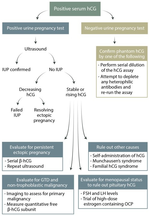 Figure 2. Diagnostic algorithm for evaluating a positive serum hCG test
(Source: Oyatogun O, et al. 2021)
References
| Target | Cat. No. | Product Name | Size | Species Reactivity | Application | Detection Sample | |
| CGB | DEIA2386 | Human hCG Visual ELISA Kit | 96T | Human | Qualitative | Serum, urine | Inquiry |
| DEIA2340 | Human B-HCG (Total) ELISA Kit | 96T | Human | Quantitative | Serum | Inquiry | |
| DEIA1895 | hCG beta ELISA Kit | 96T | Human | Quantitative | Serum, plasma | Inquiry | |
| DEIA2027 | Human HCG Beta free ELISA Kit | 96T | Human | Quantitative | Serum | Inquiry | |
| DEIA447 | hCG ELISA Kit | 96T | Human | Quantitative | Serum | Inquiry | |
| DEIA-XYA771 | hCG beta ELISA Kit | 96T | Qualitative | Cultured cells | Inquiry | ||
| DEIACL30 | CDSimple™ hCG Chemiluminescent ELISA Kit | 96T, 192T | Quantitative | Serum | Inquiry | ||
| DEIACL14 | CDSimple™ Free ?-hCG Extended Range Chemiluminescent ELISA Kit | 96T | Quantitative | Serum | Inquiry | ||
| FSHB | DEIACL27 | CDSimple™ FSH, hCG, LH & PRLs Chemiluminescent ELISA Kit | 192T, 480T, 960T | Quantitative | Serum, Plasma | Inquiry | |
| AFP | DEIACL28 | CDSimple™ AFP, hCG, uE3 Chemiluminescent ELISA Kit | 2 x 96T | Human | Quantitative | Serum | Inquiry |
| Target | Cat. No. | Product Name | Expression System | Tag/Conjugate | Application | |
| CGB | DAG-WT2648 | Human chorionic gonadotropin (HCG) control | N/A | Unconjugated | Immunoassays | Inquiry |
| DAG-WT522 | Native Human Chorionic Gonadotropin (HCG ) | Human urine | N/A | Immunogen, Control | Inquiry | |
| DAGA-776 | HCG-beta [HRP] | Human | HRP | N/A | Inquiry | |
| DAGA-775 | HCG-beta [ALP] | Human | ALP | N/A | Inquiry | |
| DAGA-774 | HCG-beta ( 95%) | Human urine | Unconjugated | N/A | Inquiry | |
| DAG-WT1087 | Native Human Chorionic Gonadotropin (hCG) | Human urine | N/A | Immunogen, Control | Inquiry | |
| DAG-WT1845 | Recombinant Human HCG alpha/beta protein | HEK293 cells | His | ELISA, CLIA, LFIA | Inquiry | |
| HCG | DAG-WT3253 | Recombinant Human HCG | CHO cells | His | ELISA | Inquiry |
| Target | Cat. No. | Product Name | Host | Isotype | Application | |
| CGB | DPABH-09232 | Rabbit anti-Human hCG beta polyclonal antibody | Rabbit | IgG | IHC-P, WB | Inquiry |
| DPABH-11274 | Anti-hCG (native) polyclonal antibody | Goat | IgG | IHC-P | Inquiry | |
| DCAB-TJ177 | Magic™ Anti-hCG beta monoclonal antibody, clone C2473N | Mouse | IgG1 | EIA, ELISA(Cap) | Inquiry | |
| DCAB-TJ178 | Magic™ Anti-hCG beta monoclonal antibody, clone C2627N | Mouse | IgG1 | EIA, ELISA(Det) | Inquiry | |
| DPABH-16425 | Rabbit anti-Human hCG beta polyclonal antibody | Rabbit | IgG | WB, ELISA, IHC-P | Inquiry | |
| HCG1995004 | DPABH-00475 | Anti-HCG1995004 (aa 113-144) polyclonal antibody | Rabbit | IgG | WB | Inquiry |
| CGA | CABT-49221MH | Anti-CGA monoclonal antibody, clone INN-hCG-45 | Mouse | IgG1 | ELISA, RIA | Inquiry |
| DCABY-4452 | Magic™ Anti-hCG monoclonal antibody, clone N29723ID2 | Mouse | IgG2b | ELISA(Cap), IA | Inquiry | |
| DCABY-4453 | Magic™ Anti-hCG monoclonal antibody, clone N82980 | Mouse | IgG1 | ELISA(Cap), WB | Inquiry | |
| DCABY-4454 | Magic™ Anti-hCG monoclonal antibody, clone NI-06 | Mouse | IgG1 | ELISA(Det), LFIA | Inquiry |
Loading ......
