
Inquiry Basket
 Loading ......
Candida albicans is a common opportunistic pathogenic fungus that colonizes human mucosal surfaces and skin. Candida albicans is usually a harmless commensal of the host. However, if changes in microbial homeostasis, immune dysfunction, or local microenvironment, Candida albicans overgrowth will give rise to infections, from superficial infections to invasive systemic candidiasis. C. albicans is the predominant pathogenic species causing candidiasis in clinical settings. It is also the most frequent cause of invasive fungal infections. It is thought that Candida albicans pathogenicity is determined by multiple properties, including adhesin proteins that mediate adherence to host cells, the ability to produce invasive hyphae and biofilm formation. Biofilms are an important survival strategy for Candida albicans, and biofilm development increases environmental fitness and causes a significant increase in drug resistance and high clinical failure rate.
A biofilm is a community of microbial cells that are attached to a solid surface or a liquid–air interface, and irreversibly embedded in a self-produced polymeric matrix. Cells in a biofilm are markedly more resistant to chemical and physical agents than their planktonic counterparts. Biofilms are prevalent in the natural environment, on artificial materials, and in body tissues. Central venous catheters, urinary catheters, pacemakers, and prostheses of joints are most commonly involved with clinical infections. The rate of biofilm infection in central venous catheters is over 50%, and most infected implanted devices need to be removed or replaced.
Biofilm formation is a dynamic and highly regulated process that can be divided into four consecutive stages. During the initial adhesion phase, yeast cells make reversible physical contact with the surface, followed by irreversible attachment mediated by various adhesins. Among these, the transcription factor Bcr1 and its downstream adhesins Als1, Als3, and Hwp1 are essential for adhesion. Once the basal layer is established, yeast cells begin to divide and trigger filamentation. Pseudohyphae and true hyphae elongate in a step-wise fashion to produce an interlaced 3D network (regulated by Efg1, Tec1 and others). Newly generated yeast cells fill up the basal layer and connect to the hyphae, and the processes of quorum sensing and contact sensing coordination of cell proliferation and morphological changes provide a robust base for 3D biofilm architecture.
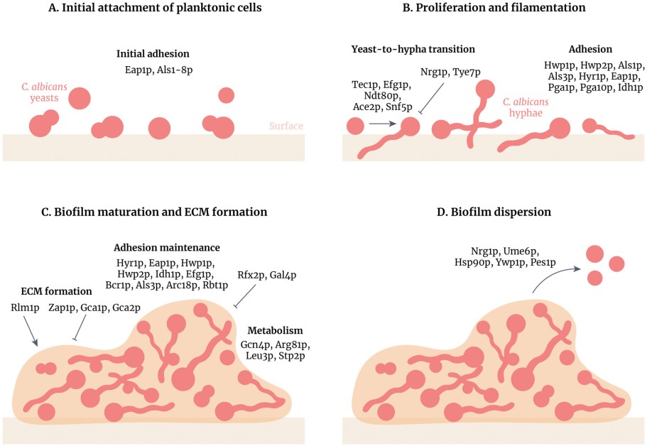 Figure 1. Stages of Candida albicans biofilm formation and development
(Source: Ponde NO, et al. 2021)
As the hyphal scaffold formation progresses from the proliferation to maturation phase, there is an increased secretion of large quantities of EPS by C. albicans. The four major classes of macromolecules that EPS is comprised of are polysaccharides, proteins, lipids, and extracellular DNA. Among these, complexes formed by α-1,6-mannan and β-1,6-glucan constitute the matrix scaffold, providing mechanical support. Although β-1,3-glucan is present in lower concentrations, it efficiently blocks antifungal drug diffusion. Additionally, the precisely arranged channel structures within EPS create an ideal microenvironment for nutrient transport and waste removal, forming functional compartmentalization between basal and apical layers. The core transcription network factors Brg1, Ndt80 and Rob1, among others, coordinately regulate the expression of about one thousand genes, required for coordinated EPS synthesis and structural remodeling. Extracellular DNA contributes to biofilm robustness by promoting cell adhesion and structural stability. Mature biofilms consistently release morphologically distinct elongated yeast cells, completing the dispersal-recolonization cycle. Surface apical hyphae produce morphologically specialized elongated yeast cells at lateral budding sites, which detach and spread through bloodstream or body fluids to new locations where they re-adhere and form secondary biofilms. Research has found that these dispersed cells exhibit significantly higher expression levels of adhesin genes (such as ALS5, ALS6, ECM33) compared to ordinary planktonic yeast, giving them enhanced colonization capacity and invasive potential on new substrate surfaces. The entire adhesion-proliferation-maturation-dispersal process typically completes within 24 to 48 hours, and the highly organized structures and robust drug resistance mechanisms exhibited in repeated cycles together establish the role of C. albicans biofilms as its major pathogenic factor.
Biofilms formation is one of the virulence factors in C. albicans pathogenesis because it not only provides fungal cells with high resistance to antifungal drugs but also results in resistance to host immune clearance, both of which are of vital importance to a dramatic increase in the intractability of the infection and the mortality of patients. In addition, clinically, C. albicans cells growing in a biofilm are typically less sensitive to the three main antifungal drug classes, that is, azoles, polyenes, and echinocandins. Biofilms are intrinsically resistant to azole drugs like fluconazole, and those formed in in vivo flow conditions show even higher tolerance. Polyene drugs are only effective against biofilms at high concentrations, but at these levels they often cause serious toxic side effects, though liposomal formulations have provided some improvement. Echinocandins target β-1,3-glucan and can effectively disrupt biofilms at therapeutic concentrations, making them the recommended first-line therapy. Biofilm drug resistance shows staged evolution involving three core mechanisms. Efflux pumps play a dominant role in early-stage biofilms: ABC transporters (Cdr1/Cdr2) and MFS family pumps (Mdr1) are significantly upregulated, actively expelling azole drugs, and knockout of related genes can restore drug sensitivity. As biofilms mature, the contribution of efflux pumps diminishes, and resistance shifts to being primarily mediated by EPS barriers. Second, matrix barriers chelate drugs (such as fluconazole) through β-1,3-glucan and eDNA, reducing local effective concentrations while physically restricting drug penetration, requiring 10–1000-fold higher concentrations to achieve minimum inhibitory concentrations. Finally, the biofilm basal layer contains persister cells representing 1–5% of the total population, a metabolically dormant yeast subpopulation that achieves intrinsic drug resistance by evading drug targets (such as the cell membrane synthesis pathways targeted by azoles).
Table 1. Classes of antifungal agents
| Antifungal drug classes | Examples | Mechanism of action | Candida albicans means of resistance |
| Azoles | Fluconazole | Inhibition of lanosterol 14 α-demethylase (ERG11; ergosterol biosynthesis) | Upregulated expression of ERG genes |
| Polyenes | Amphotericin B | Binds to ergosterol in fungal cell membranes; transmembrane pore formation, resulting in loss of membrane integrity and ion gradient disruption | Substitution of cell membrane sterols |
| Echinocandins | Caspofungin | Inhibition of β-1,3-glucan synthase | Upregulated expression of glucan biosynthesis genes |
| Pyrimidine analogs | Flucytosine | Pyrimidine analog that inhibits DNA and RNA synthesis within fungal cells | Mutations in the enzymes that catalyze the pyrimidine analog, such as FUR1 Increased synthesis of nucleotide pyrimidines that competitively inhibit the analogs |
| Allylamines | Terbinafine | Inhibition of squalene epoxidase (ERG1; ergosterol biosynthesis) | Mutations in the ERG1 gene Upregulation of drug efflux pumps |
(Source: Tsui C, et al. 2016)
The number of studies investigating the potential protective role of C. albicans biofilms in response to host immunity has been increasing over the last years. The molecules at the biofilm matrix surface differ from those at the fungal cell surface and have the potential to hinder immune recognition receptors and disrupt the functions of neutrophils, macrophages and monocytes. Biofilms, as compared to planktonic cells, were shown to inhibit the release of neutrophil extracellular trap (NET) and reduce reactive oxygen species (ROS) production. In experiments, dispersal of the matrix restored NET release. Biofilms also physically block or structurally hinder macrophage migration, resulting in decreased phagocytosis. Additionally, biofilms modulate host cytokine secretion, inducing a shift toward an ineffective Th2 immune response.
Candida albicans is an opportunistic pathogen often viewed as a single species infectious agent. Yet, it resides in the human microbiome, which is an environment that is full of different bacteria, archaea and fungi. Diet, genetics, the immune status of the host, and local environmental changes (pH, mucus viscosity, etc.) along with antibiotic administration, all shift the ecology of the microbiome as a whole and therefore susceptibility and infection course. Additionally, C. albicans is the most common fungal pathogen. In addition, C. albicans are found in polymicrobial biofilms (along with other Candida species: C. dubliniensis, C. tropicalis, C. parapsilosis, C. glabrata, etc.) in clinical patients from the oral cavity all the way to the urinary system.
In addition to those described above between members of the same genus, work has been done to understand how C. albicans coexists with some of the common bacteria it is likely to encounter and form dual species biofilms with, such as in the gastrointestinal tract, E. coli and Enterococcus faecalis. The main mechanisms by which they interact with each other are: 1) by secreting small molecule signals to control each other's behavior, such as Pseudomonas aeruginosa's 12 carbon acyl homoserine lactone that can inhibit or modify C. albicans filamentation; 2) by direct physical contact between the two cells and 3) by changing the local environment in ways such as pH or oxygen concentrations that alter each other's growth. In gut models, C. albicans biofilms can create low-oxygen "safe zones" on surfaces, supporting the growth of strict anaerobes such as Bacteroides fragilis and Clostridium perfringens even under oxygen-rich conditions. In return, these anaerobes can also reciprocally encourage C. albicans to form micro-biofilms in high O2 concentrations. This creates a mutualistic relationship with the fungi providing the bacteria with an optimal environment, while the bacteria aid in colonization and tolerance of the fungi. These findings demonstrate that fungi and bacteria create complex interactions within polymicrobial biofilms through signaling molecules, physical contact, and by altering the environment, thus changing the probability and severity of infection.
 Figure 2. Main features of C. albicans-S. aureus relationship related to biofilm lifestyle
(Source: Bernard C, et al. 2020)
No drugs have been developed that are specific to biofilms. This contributes to the problem of biofilm-associated infections being so challenging to treat. Understanding the molecular events which allow biofilms to form and be maintained could lead to new antifungal agents which target the biofilm state. An example of this is an understanding of biofilm dispersal mechanisms, which could lead to therapeutic strategies that induce early instability or inhibit the formation of correct biofilms. Clarifying the interactions between fungi and the matrix, as well as cell–cell contacts during the adhesion phase, could provide targets to block biofilm initiation or disrupt mature biofilms. Further study into the metabolic dormancy of persister cell subpopulations may help reverse their drug-resistant phenotype, thereby increasing their sensitivity to conventional treatments.
High-throughput chemical library screening has identified multiple small molecules that can inhibit biofilm formation or maintenance, but whether these compounds can be developed into clinically effective drugs remains to be evaluated. Mixing these biofilm dispersing agents with conventional antifungal agents such as fluconazole and amphotericin B may be of interest for "re-sensitizing" biofilms to their effects and to "boost" the effects of these common drugs. Natural and/or synthetic amphiphilic surfactants have also been shown to prevent biofilm development and enhance biofilm degradation from acrylic surfaces. Other biofilm development and maintenance may be inhibited by natural plant compounds such as shikonin. Thymol, an active ingredient of the herb thyme, can also trigger biofilm dispersion as well as morphological changes to in vitro biofilms in combination with fluconazole in a synergistic effect.
From the perspectives of anti-virulence and immune modulation, the antimicrobial peptides ToAP2 and NDBP-5.7 can increase fungal cell membrane permeability and induce morphological damage at both early and late biofilm stages. When combined with fluconazole or amphotericin B, they reduce effective drug concentrations and alleviate toxicity. A vaccine targeting the key adhesin Als3p (NDV-3A) significantly reduces fungal colonization in a mouse catheter implantation model, suggesting that immune prevention could also serve as a clinical adjunct.
References
| Target | Cat. No. | Product Name | Size | Species | Application | Detection Sample | |
| C. albicans | DEIA323 | Human Candida albicans IgG ELISA Kit | 96T | Human | Quantitative | Serum, plasma | Inquiry |
| DEIA324 | Human Candida albicans IgA ELISA Kit | 96T | Human | Quantitative | Serum, plasma | Inquiry | |
| DEIA325 | Human Candida albicans IgM ELISA Kit | 96T | Human | Quantitative | Serum, plasma | Inquiry | |
| DEIA809 | Candida albicans antigen ELISA Kit | 96T | Human | Quantitative, Qualitative | Serum, plasma | Inquiry |
| Target | Cat. No. | Product Name | Expression System | Tag/Conjugate | Application | |
| C. albicans | DAG-WT3855 | Purified Candida Albicans Mannan | Candida albicans | N/A | N/A | Inquiry |
| DAG-WT1977 | Inactivated C. albicans Culture Fluid | N/A | N/A | Control | Inquiry | |
| DAG-WT5417 | Native Candida albicans Unassayed Control | N/A | N/A | Immunoassays | Inquiry | |
| DAGA-553 | Candida albicans (wild strain) | C. albicans | Unconjugated | N/A | Inquiry | |
| C. albicans Enolase | DAGA-288 | Recombinant C. albicans Enolase | Sf9 insect cells | His | Inquiry | |
| DAG2702 | Recombinant Candida albicans Enolase [His] | E. coli | His | ELISA, WB, Dot | Inquiry |
Loading ......
