
 Anti-h PTH antibody
 PDF
PDF 25-OH Vitamin D3-BSA conjugated
Inquiry Basket
 Loading ......
Bone is a solid structure undergoing perpetual renewal with crucial functions such as kinematic support, visceral protection, and regulation of hematopoiesis and mineral balance. Maintenance of these functions depends on normal bone mass and strength. Skeletal diseases, such as osteoporosis, are usually accompanied by abnormal bone remodeling, in which osteoclast-mediated bone resorption preponderates over osteoblast-mediated bone formation, leading to decreased bone mass, deteriorated microstructure, and increased fragile fracture risk.
Bone needs to be remodeled to maintain integrity with osteoblasts, which are bone-forming cells, and osteoclasts, which are bone-degrading cells.
 Fig. 1: An overview of intracellular regulation of osteoclastogenesis and resorption activities
The major factors in bone formation include fibroblast growth factors (FGFs), bone morphogenetic proteins (BMPs), wingless-type (Wnt) genes, runt-related transcription factor 2 (RUNX2), and osteoblast-specific transcription factor (osterix or OSX). In addition, metabolic functions of bone largely involve the homeostasis of calcium and phosphate. Release of calcium, or absorption of calcium by bone, is largely regulated by hormones (including PTH, Calcitonin, Sex hormones, Growth hormone, and Thyroid hormones) and, less so, by steroids (Vitamin D, Glucocorticosteroids).
The maintenance of bone homeostasis is regulated by a cascade of signaling pathways. The major pathways associated with bone remodeling and pathogenesis of most bone diseases including RANKL/M-CSF/OPG, MAPKs/JNK/NF-κB, BMP2-Wnt/β-catenin, PI3K/Akt/GSK3β, and other signaling pathways. The binding of RANKL and M-CSF to their respective receptors, RANK and c-Fms, leads to upregulation of TRAF6 that activates (ERK)/JNK/MAPK signaling, NF-ĸB/nuclear factor of activated T Cells 1 (NFATc1) signaling, Ca2+ signaling, and the PI3K/Akt/GSK3β signaling pathways, leading to bone components’ degradation. The BMP-2/Wnt/β-catenin signaling pathway regulates RUNX2 activity, and BMP activates the movement of β-catenin into the nucleus to initiate osteoblast development via the canonical Wnt/β-catenin signaling pathway that elevates osteoblastogenesis (OBG) and bone production. The nuclear activity of β-catenin includes the expression of osteogenic proteins such as osteocalcin (OCN), alkaline phosphatase (ALP), and bone sialoprotein (BSP).
 Fig. 2: Role of Wnt signaling in osteoblasts
Various biomarkers are now available for specific and sensitive assessment of the rate for bone formation and bone resorption. For example, the bone formation biomarkers are total alkaline phosphatase (ALP), bone-specific alkaline phosphatase (BALP), osteocalcin (OC), procollagen type 1 N-terminal propeptide (P1NP) and procollagen type 1 C-terminal propeptide (P1CP). The bone resorption biomarkers are hydroxyproline (HYP), hydroxylysine (HYL), deoxypyridinoline (DPD), pyridinoline (PYD), bone sialoprotein (BSP), osteopontin (OP), tartrate-resistant acid phosphatase 5b (TRAP 5b), carboxy-terminal crosslinked telopeptide of type 1 collagen (CTX-1), amino-terminal crosslinked telopeptide of type 1 collagen (NTX-1) and cathepsin K (CTSK). The regulators of bone turnover are receptor activator of NF-kB ligand (RANKL), osteoprotegerin (OPG), dickkopf-1 (DDK-1) and sclerostin. These biomarkers are useful to provide the early assessment of bone diseases when the BMD measurement of DXA does not offer enough information to make the diagnosis.
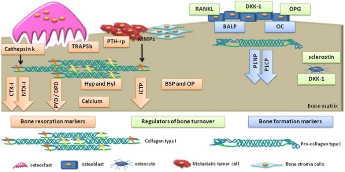 Fig. 3: Biochemical biomarkers of bone turnover
Loading ......
