
Inquiry Basket
 Loading ......
Antimitochondrial antibodies (AMA) were first discovered in the serum of PBC patients by Mackay in 1958. AMA is an antibody against the two-layer biological membrane protein of mitochondria. It is not an organ-specific or species-specific autoantibody, because mitochondrial membrane proteins are contained in the cells of various tissues and organs (liver, kidney, stomach, heart).
Mitochondrial antigens are located in the mitochondrial membrane of eukaryotic cells. The antigen components are subunits of the 2-oxoacid dehydrogenase complex and are a group of autoantigens. AMA target antigens are divided into 9 types (M1-M9). M1 is cardiolipin in the outer mitochondrial membrane; M2 is the main component of the AMA reaction in the serum of PBC patients. Its essence is Pyruvate dehydrogenase and alpha-keto acid dehydrogenase complex on the inner mitochondrial membrane; the nature of M3 is unclear; M4 is sulfite oxidase; M5 is a 65kD protein; the properties of M6, M7, and M8 are unknown; M9 is a glycogen phosphorylase.
Due to the complex composition of mitochondrial antigens, the properties of many antigens are not yet understood. According to the antigens targeted, mitochondrial antibodies are divided into nine subtypes, namely anti-M1, anti-M2, anti-M7 for the inner membrane and anti-M7, Anti-M3, anti-M4, anti-M5, anti-M6, anti-M8, anti-M9 for the outer membrane, each with different characteristics.
Anti-M1 is anticardiolipin antibody, which is not currently included in anti-mitochondrial antibodies; anti-M2, M4, M8, and M9 antibodies are mainly related to primary biliary cirrhosis (PBC) PBC, especially anti-M2, has a very high specificity for PBC and is found in 90% of PBC patients. It is often used as an important laboratory diagnostic indicator of the disease, but it has no correlation with the stage, disease severity and treatment effect of PBC. Except for PBC, anti-M2 Also seen in chronic active hepatitis, HBsAg-negative liver disease, etc.; anti-M3 is seen in patients with drug-induced lupus erythematosus (PLE) syndrome induced by the pyrazolone series of drugs; anti-M5 is seen in SLE and autoimmune hemolytic anemia; M6 is seen in hepatitis induced by iproniazid (a monoamine oxidase inhibitor); anti-M7 appears in some patients with cardiomyopathy of unknown origin. Its target antigen is organ-specific and exists in the mitochondria of cardiomyocytes.
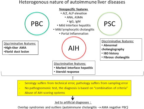 Figure 1. Heterogenous nature of autoimmune liver diseases. (Ozaslan E, et al.; 2016)
IFA is the main screening method for detecting AMA. When AMA is positive, the immunostrip method or ELISA method using purified antigen as the coated antigen must be used for mitochondrial antibody typing.
The indirect immunofluorescence method using rat (or monkey) liver, HEp-2 cells or rat kidney as the reaction matrix is usually used for detection. The AMA in the specimen will bind to the mitochondrial components of these tissues or cells, and then specifically bind to the antibody molecules bound to the tissues or cells through the fluorescein-labeled antibody. After washing the unbound fluorescent secondary antibodies, place the substrate piece. Observed under a fluorescence microscope, the presence of a characteristic fluorescence pattern is considered positive.
Under a fluorescence microscope, when AMA is positive in the specimen to be tested, granular fluorescence from fine to coarse will appear in the cell cytoplasm. In HEp-2 cells, there is coarse granular fluorescence in the cytoplasm. In renal tissue sections, the proximal renal tubules of AMA-6 (M6) and AMA-9 (M9) have strong fluorescence, while the distal renal tubules are negative. The distal renal tubules of AMA-M7 and AMA-M8 are fluorescent, while the proximal renal tubules are negative. The proximal and distal renal tubules of other types of AMA show obvious fluorescence, and the glomerular fluorescence is weak. In gastric tissue sections, parietal cells were obviously fluorescent, while chief cells were weakly fluorescent. In liver tissue sections, the liver cytoplasm showed fine sand-like fluorescence.
Highly purified mitochondrial antigens such as M2 (pyruvate dehydrogenase complex extracted and purified from pig heart), M4 (sulfite oxidase extracted and purified from chicken liver), M9 (glycogen phosphate purified from rabbit muscle) enzyme) were respectively coated on the microplate, blocked and incubated with the patient specimen. If the specimen contains AMA, it will bind to the M2, M4 or M9 antigen on the solid phase, and then add human enzyme-labeled anti-human IgG (anti-M9 The detection uses enzyme-labeled anti-human IgG and IgM warm antibodies) and enzyme substrate to develop color, and the concentration of anti-M2, anti-M4, or anti-M9 antibodies is determined according to the degree of color development.
Result judgment: (1) Qualitative detection: if the color development degree is lower than the cut-off value, it is negative; if it is higher than the cut-off value, it is positive. (2) Quantitative test: Create a standard curve with the concentration of AMA-related standards as the abscissa and the corresponding absorbance value as the ordinate. The corresponding AMA concentration can be found according to the standard curve.
Purified antigens of M2 (pyruvate dehydrogenase complex), M4 (sulfite oxidase), and M9 (glycogen phosphorylase) were coated on membrane strips in parallel as solid-phase antigens. The AMA in the specimen specifically binds to the purified antigen on the membrane strip to form an immune complex, which is washed to remove non-specific binders, and then human enzyme-labeled anti-human IgG antibody (or anti-human IgG, IgA, IgM) is added, and the enzyme-labeled secondary antibody binds to the strip antigen. The antibody specifically binds, washes away the unbound enzyme-labeled secondary antibody, and then adds the substrate. The enzyme catalyzes the substrate to generate an insoluble chromogenic product to develop the band. The results of different AMAs can be judged based on the type and color of the antigen on the membrane strip.
If the antigen band is colorless, it is negative, if it is vaguely visible, it is suspicious, and if it is obviously colored, it is positive. The degree of positivity can be estimated based on color detection.
References
| Target | Cat. No. | Product Name | Expression System | Tag/Conjugate | Application | |
| Mitochondrion | DAG-T1243 | Mitochondrial Antigen | Inquiry |
| Target | Cat. No. | Product Name | Size | Species Reactivity | Application | Detection Sample | |
| M2 type mitochondria | DEIA1224 | Human M2 type mitochondria Antibody ELISA Kit | 96T | Human | Quantitative | culture supernatants, serum, plasma, tissues | Inquiry |
| Mitochondria | DEIA1904 | Mitochondria ELISA Kit | 96T | Human | Qualitative | serum | Inquiry |
| ROS1 | DEIA-LL203 | Human ROS1 ELISA Kit | 96T | Human | Quantitative | Serum, plasma, tissue homogenates and other biological fluids | Inquiry |
| AMA-M2 | DEIA1826 | AMA-M2 Antibody ELISA Kit | 96T | Human | Quantitative | Serum, plasma | Inquiry |
| DEIA3129 | AMA-M2 Ab ELISA Kit | 96T | Human | Quantitative | Serum, plasma | Inquiry |
Loading ......
