
Inquiry Basket
 Loading ......
The researchers initially found that antigens in salivary and lacrimal gland extracts from patients with Sjögren's syndrome (SS) reacted specifically with two precipitating antibodies, called SjD and SjT, respectively. Among them, the SjD antigen is insensitive to trypsin or heat, while the SjT antigen can be destroyed by the same treatments. Subsequently, as the study progressed, the investigators found the same phenomenon in various tissue extracts from patients with systemic lupus erythematosus (SLE) in addition to patients with SS, and named the cytoplasmic antigen Ro and the cytosolic antigens SSA and SSB, both characterized by resistance to RNase and DNase, and sensitivity to trypsin was determined by the individual antigens. Subsequently, as Ro and La were shown to be antigenically identical to SSA and SSB, these antigenic systems became known as Ro/SSA and La/SSB.
Anti-Ro/SSA autoantibodies are the most common nuclear antibodies found in the sera of patients with connective tissue diseases. The Ro antigen consists of two different Ro proteins (52kD and 60kD), of which Ro60 binds to hY-RNAs. Ro60 belongs to the TROVE family of proteins, encoded by a 1.8 kb gene on chromosome 19, and is involved in the repair of misfolded non-coding RNAs and cell survival in response to UV irradiation. Ro52 is encoded by a gene on chromosome 11 with zinc-finger and leucine-zipper domains and is a TRIM-family E3 ligase that regulates the production of pro-inflammatory cytokines, induction of apoptosis, and antiviral responses. Anti-Ro antibodies of different specificities are associated with different clinical manifestations, and the detection of anti-Ro/SSA specificity in joints, kidneys, skin, and lungs, respectively, is prognostically useful. In addition, the pathogenesis of neonatal lupus erythematosus (NLE) is closely related to anti-SSA/Ro52. Ro52 and Ro60 have different conformational dependencies for their epitopes. Most sera in SSA/Ro52 usually recognize linear epitopes located at the leucine zipper site, whereas SSA/Ro60 recognizes conformational epitopes, and once protein denaturation occurs, antibody binding to Ro60 also disappears.
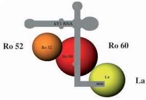 Figure 1. The Ro/La RNA complex
(Source: Franceschini F, et al. 2005)
The expression of anti-SSA/Ro antibodies is primarily associated with connective tissue diseases (CTDs), and the most common disease diagnoses in subjects with positive expression are lupus (mostly systemic lupus erythematosus), rheumatoid arthritis (RA), SS, systemic sclerosis, and dermatomyositis, as well as in mothers of neonatal lupus-affected children. Commonly used laboratory methods for the clinical detection of anti-Ro/SSA antibodies include recombinant Ro antigen-based ELISA, FEIA, and line-blot immunoassay (LIA). In particular, traditional anti-SSA/Ro assays are more biased toward Ro60 and some anti-SSA/Ro52 positive serum precipitates are negative, undetectable by natural SSA/Ro-based ELISA, and do not have specific ANA fluorescent staining patterns. As the assays have evolved, investigators have found that immuno-WB assays using natural Ro antigens are more sensitive and can detect anti-Ro/SSA positivity in the general population as well as arrhythmogenic anti-SSA/Ro52 autoantibodies in autoimmune congenital heart block (aCHB).
Detection of a positive anti-SSA/Ro52 antibody does not directly diagnose the disease. For example, one study found that two patients who tested positive for anti-SSA/Ro52 were infected with the hepatitis C virus but were not diagnosed with SS. Using three different laboratory methods, the frequency of anti-Ro52 antibodies was similar to the frequency of anti-Ro60 in the other disease types except myositis and systemic sclerosis. On the other hand, the percentage of those with anti-Ro52 antibodies without anti-Ro60 antibodies varied from 5.4% in the pediatric SLE group to 35.4% in the myositis group. In the SS group, Ro60 autoantibodies were also present in 63.2% of anti-Ro52 sera.
Anti-SSA/Ro associated long-QT syndrome
In the last century, it was recognized that mothers with autoimmune diseases could deliver autoantibodies across the placenta to the fetus, resulting in a fetus with congenital complete atrioventricular block. It was found that neonates/infants without autoimmune congenital heart block born to anti-Ro/SSA positive mothers had a longer QTc than anti-Ro/SSA negative controls. However, this phenomenon of disturbance of ventricular repolarization is functional and reversible and returns to normal spontaneously in the first year of life when the anti-SSA/Ro antibodies of maternal origin disappear.
Similarly, QTc prolongation and increased incidence of VAs have been reported in circulating anti-Ro/SSA antibody-positive adults. QTc prolongation (>440 ms in 45-60% of cases) is frequently observed in anti-Ro/SSA-positive adults with CTD, occurring 5-12 times more frequently than in normal subjects, and is associated with autoantibody levels. A detailed study showed that only the serum concentration of the anti-Ro/SSA-52kD isoform was significantly associated with QTc time.
Despite the data from many studies demonstrating a clinical association between anti-Ro/SSA antibodies and the risk of LQTS, many studies in children or adults have reported conflicting results. The reasons for this are analyzed as follows. First, the circulating levels of anti-Ro/SSA-52kD autoantibodies in patients may not be sufficient to induce measurable electrocardiographic changes. In fact, the concentration of anti-Ro/SSA-52kD varies considerably among CTDs, and specific subtypes were not evaluated in most of the negative correlation studies. Second, most of the studies were retrospective, with different thresholds for defining QTc prolongation, which may have led to inconsistent results. Defining prolongation too long may result in too few cases with QTc prolongation and an insufficient sample size for statistical comparisons. Considering only qualitative data on anti-Ro/SSA positivity without considering the subtypes and their concentrations can also lead to a lack of scientific validity in data analysis. Finally, anti-Ro/SSA antibodies can inhibit multiple cardiac ion channels simultaneously, which can have conflicting effects on action potentials and, consequently, on the QT intervals detected by ECG. These multiple effects on cardiomyocyte electrophysiology, as well as individual differences in ion channel reserve, may also contribute to the wide variability in clinical outcomes.
The QT-prolonging effect of anti-Ro/SSA antibody (especially the anti-Ro/SSA-52kD subtype) is due to the inhibition of the associated current IKr due to the specific cross-reaction with the cardiac hERG-K+ channel. The direct electrophysiological nature of this effect explains why circulating anti-Ro/SSA antibodies are associated with an increased risk of QTc prolongation/TdP in the clinical setting.
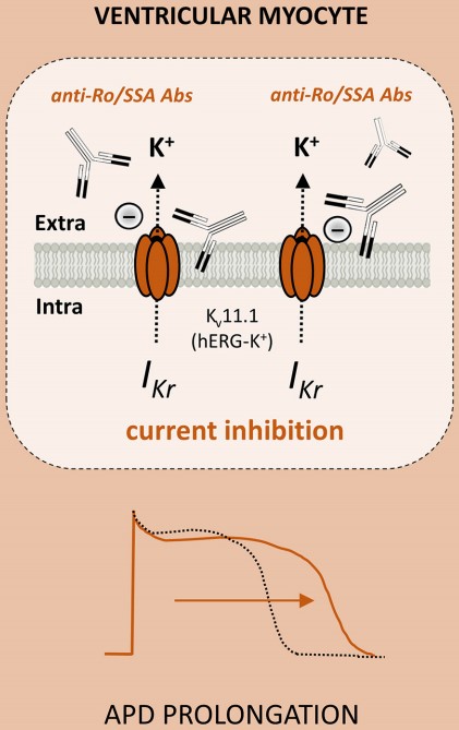 Figure 2. Anti-Ro/SSA antibodies inhibit the IKr current by directly recognizing hERG potassium channel
(Source: Lazzerini PE, et al. 2021)
Anti-SSA/Ro associated cardiac rhythm disturbances
Anti-Ro/SSA mediates atrioventricular (AV) and sinoatrial (SA) nodal conduction disturbances, the causes of which are as follows. The action potential of the cardiac conduction system is highly dependent on calcium channel currents. The serum and immunoglobulin G of Ro/SSA-positive patients can bind to two types of calcium channels (L-type (long-acting) calcium channel and T-type (transient) calcium channel), down-regulate them, and disrupt intracellular calcium homeostasis, resulting in apoptosis. Not all antibodies cross-react with calcium channels, and anti-Ro/SSA-52 antibodies are effective, but patients with positive anti-Ro/SSA-60 antibodies are less likely to develop this complication. Secondly, it can induce apoptosis. In the process of apoptosis, Ro52 will be redistributed from the cytoplasm to the cell membrane so that it can be recognized by the immune system and autoantibodies will be synthesized against it. In addition, anti-Ro/SSA-52 antibodies with apoptotic cellular immune complexes can induce type 1 interferon. Finally, anti-Ro/SSA can induce fibrosis. Macrophages can bind to conditioned cardiomyocytes via their FcR. The co-culture of macrophages and conditioned cardiomyocytes showed the characteristics of promoting inflammation, and the cultured supernatant could induce cardiomyocytes to differentiate into fibroblasts associated with fibrosis.
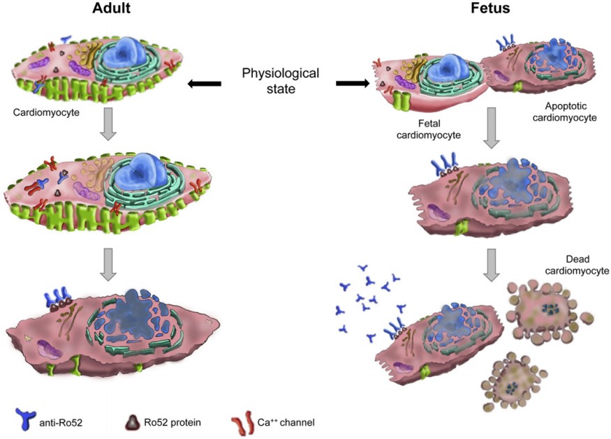 Figure 3. Induction of apoptosis
(Source: Santos-Pardo I, et al. 2015)
Anti-SSA/Ro associated autoimmune liver disease
Anti-Ro/SSA52 specific antibodies are frequently seen in autoimmune liver diseases. It is the most common anti-ENA response in primary biliary cirrhosis, and diagnosis is associated with elevated serum bilirubin and IgM levels. With further study and follow-up, anti-SSA/Ro52 antibodies may identify patients with primary biliary cirrhosis in advanced and active stages of the disease. Serum and soluble liver antigen (anti-SLA) autoantibodies in autoimmune hepatitis I (AIH I) react simultaneously with the Ro52 autoantigen. Anti-SLA autoantibodies are highly specific for AIH I and recognize a single antigen, UGA tRNA suppressor-associated protein (tRNP(Ser)Sec). 98% of anti-SLA-positive serum samples react with SSA/Ro52, and compared to tRNP(Ser)Sec-negative patients with AIH I, these patients have more severe biochemical and histologic disease.
Anti-SSA/Ro associated SLE and SS
Anti-Ro antibodies are commonly associated with SLE, SS/SLE overlap syndrome, subacute cutaneous lupus erythematosus (SCLE), and NLE. Anti-Ro antibodies are positive in 70%-100% and 40%-90% of patients with SS and SLE, respectively. Patients with C2- and C4-deficient SLE tend to have anti-Ro antibodies with cutaneous symptoms and polyarthritis, but without renal or CNS features. Anti-Ro antibodies appear earlier than other autoantibodies associated with SLE, on average 3 or 4 years before diagnosis. The presence of anti-Ro antibodies in the tears of SS patients correlates with the severity of keratoconjunctivitis sicca.
References
| Target | Cat. No. | Product Name | Size | Species Reactivity | Application | Detection Sample | |
| TRIM21 | DEIA1703 | Human SSA IgG ELISA Kit | 96T | Human | Semi-quantitative | Serum | Inquiry |
| DEIA1220 | Human SSA Antibody ELISA Kit | 96T | Human | Qualitative | Cell culture supernatant, serum, plasma, tissues | Inquiry | |
| DEIA6284 | SS-A/Ro Ab/SS-A/Ro Ak ELISA Kit | 96T | Human | Quantitative, Qualitative | Serum | Inquiry | |
| DEIA4222 | SS-A (Ro) IgG ELISA Kit | 96T | Human | Quantitative | Serum, plasma | Inquiry | |
| DEIA4225 | SS-A Ab ELISA Kit | 96T | Human | Quantitative | Serum, plasma | Inquiry | |
| DEIANS078 | Human anti-SSA/Ro antibody ELISA Kit | 96T | Human | Qualitative | Serum, plasma | Inquiry | |
| DEIANS077 | SS-A/Ro-Ab ELISA Kit | 96T | Human | Quantitative, qualitative | Serum or plasma (EDTA, citrate, heparin). | Inquiry |
| Target | Cat. No. | Product Name | Host | Isotype | Application | |
| TRIM21 | DPABH-13922 | Anti-TRIM21 (aa 12-262) polyclonal antibody | Rabbit | IgG | WB, IHC-P | Inquiry |
| DCABH-13830 | Anti-TRIM21 monoclonal antibody | Rabbit | IgG | WB | Inquiry | |
| DPAB-DC2965 | Anti-TRIM21 (aa 68-175) polyclonal antibody | Mouse | WB, ELISA | Inquiry | ||
| CABT-B11670 | Mouse anti-Human TRIM21 monoclonal antibody, clone 3D0 | Mouse | IgG2a | WB, IHC, sELISA, ELISA | Inquiry | |
| DMAB-JX2362040 | Rabbit Anti-Human TRIM21 Monoclonal Antibody, Clone S12-2F9 | Rabbit | IgG | WB | Inquiry | |
| CABT-L6300 | Human Anti-Human Ro52 monoclonal antibody, clone D42 | Human | IgG | ELISA | Inquiry |
| Target | Cat. No. | Product Name | Expression System | Tag/Conjugate | Application | |
| TRIM21 | DAG-T1225 | Recombinant Ro52 Antigen | Inquiry | |||
| CDBP2816 | Human TRIM21 blocking peptide | N/A | Unconjugated | Apuri, BL, ELISA | Inquiry | |
| TRIM21 | DAG-WT2282 | Recombinant Human SS-A/Ro 52 [His] | E. coli | His | Immunoassays | Inquiry |
| Target | Cat. No. | Product Name | Size | Species Reactivity | Application | Detection Sample | |
| TROVE 2 | DEIA114J | Mouse Anti-SSA (Ro-60) ELISA Kit | 96T | Mouse | Quantitative | Serum | Inquiry |
| DEIA103J | Anti-SSA (Ro-60) ELISA Kit | 96T | Human | Quantitative | Serum | Inquiry |
| Target | Cat. No. | Product Name | Host | Isotype | Application | |
| TROVE 2 | CABT-L6299 | Human Anti-Human SSA monoclonal antibody, clone D41 | Human | IgG | ELISA | Inquiry |
| DPABHZ04 | Anti-Ro/SS-A polyclonal antibody | Human | ELISA | Inquiry | ||
| DPABH-24658 | Anti-TROVE2 (aa 466-495) polyclonal antibody | Rabbit | IgG | WB | Inquiry | |
| DPABH-25683 | Anti-TROVE2 (aa 1-50) polyclonal antibody | Rabbit | IgG | WB, IP | Inquiry | |
| DCABH-4115 | Anti-TROVE2 monoclonal antibody, clone 3C5 | Mouse | IgG2a | WB, ELISA, IHC-P | Inquiry | |
| DPAB-DC2966 | Anti-TROVE2 (aa 1-100) polyclonal antibody | Mouse | WB, ELISA | Inquiry | ||
| DPABH-19482 | Anti-TROVE2 (aa 178-429) polyclonal antibody | Rabbit | IgG | WB | Inquiry |
| Target | Cat. No. | Product Name | Expression System | Tag/Conjugate | Application | |
| TROVE 2 | DAG-T1224 | Ro (SSA) Antigen | Inquiry | |||
| DAGC113 | Recombinant Human Ro/SS-A (60 KDa) Protein [His, biotin] | E. coli | His, biotin | SDS-PAGE, ELISA | Inquiry | |
| DAG4849 | Human Ro/SS-A [His] (60 kDa) | E. coli | His | WB, ELISA | Inquiry | |
| DAG4851 | Human Ro/SS-A [His] (52 kDa) | Insect cells | His | WB, ELISA | Inquiry | |
| DAGC115 | Recombinant Human Ro/SS-A (52 KDa) Protein [His, biotin] | Insect Cells | His, biotin | ELISA | Inquiry | |
| DAG-T1224 | Ro (SSA) Antigen | Inquiry |
Loading ......
