Dengue is a viral illness that is spread primarily by Aedes mosquitoes and infects ~390 million people each year worldwide. It is caused by dengue virus (DENV), which belongs to the family of Flaviviridae in the genus Flavivirus. The DENV particle consists of a single-stranded positive-sense RNA enclosed within a capsid that is surrounded by an envelope containing the proteins E and prM/M. The three structural proteins (capsid, E and prM/M) plus seven non-structural proteins are transcribed and translated during viral replication, making them accessible to antigen-processing pathways within the cell, and the genome is synthesized through a double-stranded RNA intermediate. Adaptive immunity, involving multiple subclasses of antibodies and various subsets of B and T cells, can aid in the resolution of primary infection and prevent symptomatic infection by homologous DENV strains during secondary infection. The induction of such responses against all four DENV serotypes is the prime aim of vaccination.
View All Dengue Virus Antigens
View All Dengue Virus ELISA Kits
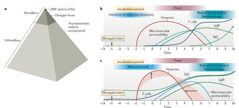 Fig1. Time course of acute infection and immune responses during symptomatic dengue virus infection.
(Source: Nature Reviews Immunology, 2019)
Dengue immunity at the site of infection. At the site of the mosquito bite in the skin, various immune sentinels such as dendritic cells (DCs), Langerhans cells (LCs), macrophages and mast cells (MCs) are present. Some skin cells, including DCs and macrophages, are known DENV infection target cell types, and either productive or abortive infection by DENV can trigger antiviral innate immune responses through the activation of pattern recognition receptors. MCs are granulated immune cells that are preloaded with immune mediators and can respond to certain stimuli, including DENV, with a degranulation response within minutes.
Initiation of dengue virus- specific adaptive immune responses. DENV infection starts in the human skin, yet the virus must achieve viraemia in order to infect new vector hosts for its spread. To accomplish this, DENV is lymphotropic. Studies in primates and mice have shown that DENV targets skin-draining LNs and infects cells that traffic through them, such as DCs and monocytes, to establish systemic infection. Although infection proceeds through LNs, the functionality of these organs in generating specific immunity is apparently not substantially compromised because adaptive immunity to DENV is largely protective against viruses of the same serotype. Despite the potential of DCs to become infected, they are also critical for antigen presentation to T cells in LNs, and there is evidence that the processing and presentation of DENV antigens influences downstream disease outcomes.
Immune clearance of DENV in vivo is coordinated by multiple cell types and mechanisms. These include innate immune responses (such as production of type I and type II interferons) by the cells directly infected with DENV, cell killing by cytotoxic lymphocytes and production of neutralizing antibodies by B cells.
New insights into protective CD4+ T cell responses to dengue virus. The control of DENV infection in asymptomatic individuals correlates with increases in numbers of activated CD4+ T cells and is characterized by a transcriptional response of peripheral blood mononuclear cells that is indicative of antigen presentation and T cell activation. Moreover, certain CD4-restricted HLAs, such as HLA-DRB1, have been linked to less severe DENV infection outcomes, suggesting that CD4+ T cells have a protective role.
CD8+ T cell responses during primary dengue infection. For patients with acute primary DENV infection, CD8+ T cell responses peak around the day of defervescence or slightly before, as shown by DENV-specific tetramer staining. Immune memory also persists following contraction of the T cell response. During the febrile stage of illness, a massive expansion of activated CD8+ T (CD38+HLA-DR+) cells is observed. It is also noteworthy that high levels of both IFNα and IFNγ were found in the plasma of patients during early primary DENV infection, suggesting that cells other than T cells were the major source of IFNγ.
Epitopes for CD4+ and CD8+ T cell recognition. Views on the functional influence of T cell responses in DENV infection may lack consensus, but efforts to identify T cell epitopes are likely to clarify which virus-directed immune responses are protective and whether any are also harmful. Many studies highlight T cell epitopes that may be widely recognized in the human population and indicate that both non-structural and structural proteins can contain immunodominant CD4+ and CD8+ T cell epitopes.
B cells and antibody responses after primary infection. Within days following infection, plasma blasts and titers of DENV-specific antibodies increase in the blood. DENV-specific B cells were highly serotype-specific in primary infection.
Reference
| Target | Cat. No. | Product Name | Size | Species Reactivity | Application | Detection Sample | |
| DENV | DEIABL333 | Dengue virus IgM µ-capture ELISA Kit | 96T | Qualitative | serum, plasma | Inquiry | |
| DEIA508 | Dengue IgG ELISA Kit | 96T | Human | Qualitative | serum, citrate plasma | Inquiry | |
| DEIA510 | Dengue IgM Capture ELISA Kit | 96T | Human | Qualitative | serum | Inquiry | |
| DEIA1439 | Dengue Virus IgG Human ELISA Kit | 96T | Human | Qualitative | serum, citrate plasma | Inquiry | |
| DENV NS1 | DEIABL14 | Dengue NS1 Antigen ELISA Kit | 96T | Human | Qualitative | Human Serum | Inquiry |