Antigen Description
Neurofilaments are the 10 nanometer (10nm) intermediate filaments found specifically in neurons. They are a major component of the cell"s cytoskeleton, and provide support for normal axonal radial growth (i.e. increases in axon"s diameter). Neurofilaments are composed of polypeptide chains or subunits that are related structurally to the intermediate filaments of other tissues such as keratin subunits, which make 10nm filaments expressed specifically in epithelia. The family of proteins making intermediate filaments is divided into 5 major classes, the keratins forming the classes I and II. Class III contains the proteins Vimentin, Desmin, Peripherin and Glial Fibrillary Acidic Protein (GFAP). The major neurofilament subunits occupy the class IV family of intermediate filaments. Finally class V contains the nuclear lamins.
Temporal progression of tau pathology and neuroinflammation in a rhesus monkey model of Alzheimer's disease
Beckman D, Diniz GB, Ott S, Hobson B, Chaudhari AJ, Muller S, Chu Y, Takano A, Schwarz AJ, Yeh CL, McQuade P, Chakrabarty P, Kanaan NM, Quinton MS, Simen AA, Kordower JH, Morrison JH
Applications: IF
Reactive species: Rhesus macaque
"Abstract: Introduction:The understanding of the pathological events in Alzheimer's disease (AD) has advanced dramatically, but the successful translation from rodent models into efficient human therapies is still problematic.
Methods:To examine how tau pathology can develop in the primate brain, we injected 12 macaques with a dual tau mutation (P301L/S320F) into the entorhinal cortex (ERC). An investigation was performed using high-resolution microscopy, magnetic resonance imaging (MRI), positron emission tomography (PET), and fluid biomarkers to determine the temporal progression of the pathology 3 and 6 months after the injection.
Results:Using quantitative microscopy targeting markers for neurodegeneration and neuroinflammation, as well as fluid and imaging biomarkers, we detailed the progression of misfolded tau spreading and the consequential inflammatory response induced by glial cells.
Discussion:By combining the analysis of several in vivo biomarkers with extensive brain microscopy analysis, we described the initial steps of misfolded tau spreading and neuroinflammation in a monkey model highly translatable to AD patients.
Highlights:Dual tau mutation delivery in the entorhinal cortex induces progressive tau pathology in rhesus macaques. Exogenous human 4R-tau coaptates monkey 3R-tau during transneuronal spread, in a prion-like manner. Neuroinflammatory response is coordinated by microglia and astrocytes in response to tau pathology, with microglia targeting early tau pathology, while astrocytes engaged later in the progression, coincident with neuronal death. Monthly collection of CSF and plasma revealed a profile of changes in several AD core biomarkers, reflective of neurodegeneration and neuroinflammation as early as 1 month after injection."
Article snippet: Neuronal markers: Pan‐Neurofilament (1:400; DMAB 7133, Creative Diagnostics, New York, NY) 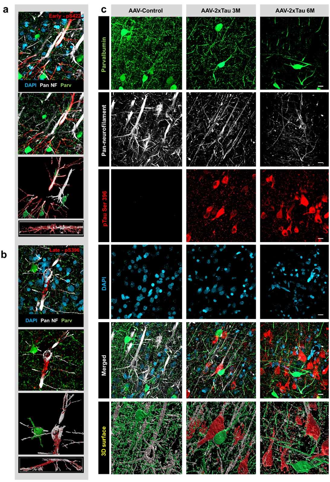 Figure 1. Virally-induced Tau overexpression correlates with cytoskeletal breakdown in large pyramidal neurons.
  Figure 2. Activated microglia is associated with synaptic and neuronal loss in the HF-ERC area.

 COA
COA Publication


 Loading ......

