
Inquiry Basket
 Loading ......
Measles virus is the causative agent of measles and belongs to the genus Morbillivirus of the family paramyxoviridae. Measles is a common acute infectious disease in children. It is highly contagious and is characterized by rash, fever and respiratory symptoms. If there are no complications, the prognosis is good. Studies have found that subacute sclerosing panencephalitis (SSPE) is related to the measles virus. Additionally, measles infection can damage and suppress the entire immune system. This means that people who have had measles are more susceptible to other infectious diseases. The effects can last up to three years after they recover from measles.
Measles virus is spherical or filamentous, with a diameter of about 120nm~250nm. The core is single negative-strand RNA, which is not segmented. The full genome length is about 16kb. The genome has 6 genes, N, P, M, F, H, and L, respectively. Encodes 6 structural and functional proteins: nucleoprotein (NP), phosphoprotein (P), M protein (membrane protein, M), fusion protein (F), hemagglutinin, H) and RNA-dependent RNA polymerase (large polymerase, L). The nucleocapsid has a helical symmetry, an envelope on the outside, and two spikes on the surface, namely hyaluronic acid (HA) and hemolyxin(HL). Their components are both glycoproteins, but their properties are different. HA can only agglutinate monkey red blood cells and can also adsorb to host cell receptors. HL has the function of hemolysis and cell fusion to form multinucleated giant cells. Both HA and HL have antigenicity, and the corresponding antibodies produced have protective effects.
 Figure 1. Schematic representation of measles virus.(Aref S, et al.; 2016)
The only natural reservoir of measles virus is humans. Patients in the acute phase are the source of infection, and patients are infectious from 6 days before to 3 days after the onset of rash. It is spread through droplets and can also be spread through utensils, toys or close contact. Measles is highly contagious, and almost all susceptible people will become ill after exposure. The incubation period of onset is 9 to 12 days. Since CD46 is a measles virus receptor, most tissue cells with CD46 can be target cells for measles virus infection. The virus entering through the respiratory tract first binds to the receptors of respiratory epithelial cells and proliferates therein, then invades the lymph nodes and proliferates, and then enters the blood (proliferates well in white blood cells), forming the first viremia. The virus reaches the lymphoid tissues of the body and proliferates in large quantities and then enters the blood again, forming a second viremia. Fever begins at this time, followed by upper respiratory tract catarrhal symptoms due to virus proliferation in the conjunctiva, nasopharyngeal mucosa, and respiratory mucosa. The virus also multiplied in the dermis, and Koplik spots with gray center and red surroundings appeared on the inner mucosa of the oral cheeks. A characteristic rash appeared 3 days later. The main cause of the rash was local hypersensitivity reaction. Generally, 24 hours after the rash appears in children, the body temperature begins to drop, the respiratory symptoms subside in about a week, and the rash becomes darker and has pigmentation. Some young and frail children are prone to bacterial infections, such as secondary bronchitis and otitis media, and are especially susceptible to bacterial pneumonia, which is the main cause of death in children with measles. About 0.1% of patients develop encephalomyelitis. It is a delayed-type hypersensitivity disease that often occurs one week after recovery. It shows typical pathological changes of demyelination and obvious lymphocyte infiltration, and often remains. There are permanent sequelae, and the mortality rate is 15%. Immunocompromised children infected with measles virus often do not have rash, but can develop severe and fatal measles giant cell pneumonia. One in a million measles patients develops subacute sclerosing panencephalitis (SSPE) several years after recovery, often before school age. SSPE is a delayed complication of acute infection, manifesting as progressive brain decline and death within 1 to 2 years. Studies have found that although there are high-titer IgG or IgM anti-measles virus antibodies in the patient's serum and cerebrospinal fluid, it is difficult to isolate the measles virus using these antibodies. It is now believed that the virus in brain tissue is a measles-deficient virus. Due to the mutation of the viral M gene in brain cells, it lacks the ability to synthesize the measles virus M protein, thus affecting the assembly, budding and release of the virus. Therefore, measles virus can be isolated by co-culturing SSPE autopsy brain tissue cells with measles virus-sensitive cells (such as HeLa, Vero, etc.).
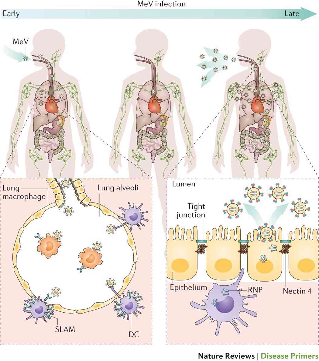 Figure 2. Measles virus infection and transmission.(Rota PA, et al.; 2016)
After measles disease, the human body can acquire lifelong immunity, which mainly includes humoral immunity and cellular immunity, with cellular immunity playing a major role. Both anti-HA antibodies and HL antibodies produced after infection can neutralize the virus, and HL antibodies can also prevent the virus from spreading between cells. IgM is dominant in the early stage of infection, and then IgG1 and IgG4 are dominant. Cellular immunity has a strong protective effect. For example, people with immunoglobulin deficiency can recover from measles and resist reinfection; while people with cellular immunity deficiency have extremely severe measles infections, which shows that cellular immunity plays a leading role in the recovery of the body. Specific killer T cells can be detected in the peripheral blood at the early stage of the rash. Babies under 6 months old are less susceptible to infection because they acquire IgG antibodies from their mothers. However, as they age, the antibodies gradually disappear, their autoimmunity is not yet complete, and their susceptibility increases. Therefore, measles is more common in infants and young children between 6 months and 5 years old.
Typical measles cases do not require laboratory testing and can be diagnosed based on clinical symptoms. For mild and atypical cases, microbiological examination is required to confirm the diagnosis. Since virus isolation and identification methods are complex and time-consuming, requiring at least 2 to 3 weeks, serological diagnosis is often used.
Virus Isolation
Although there are currently many types of laboratory detection methods for measles virus, virus isolation is still an irreplaceable classic method, which is mostly used for molecular epidemiological research on viruses.
The patient's blood, throat washing fluid or throat swabs in the early stage of the disease are taken and treated with antibiotics, and then inoculated into human embryonic kidney, monkey kidney or human amniotic membrane cells for culture. The virus proliferates slowly, and typical CPE may appear after 7 to 10 days, that is, multinucleated giant cells with eosinophilic inclusions in the cells and nuclei. Immunofluorescence technology is then used to confirm the measles virus antigen in the inoculated culture.
Virus isolation methods have been gradually replaced by some fast, accurate and simple detection methods due to long culture cycles, complex experimental operations, time constraints, and many biological influencing factors. Despite this, virus isolation remains the classic method for virus detection. Because viruses mutate quickly, virus isolation methods can isolate the latest popular virus mutant strains, which is of great significance for identifying new infectious diseases and can also isolate strains for vaccine development.
Serological Diagnosis
There are many serological methods used for MV detection. In the early stage, there were hemagglutination inhibition (HI) test, neutralizing antibody test (NT), complement fixation test (CF), etc., which can be used for the detection of viruses and virus antibodies and the isolation and identification of viruses, especially HI and NT are still important methods for MV identification and diagnosis, playing an important role in detection, isolation and identification.
Immunological Testing
Enzyme-linked immunosorbent assay (ELISA), immunofluorescence assay (IFA), solid-phase radioimmunoassay (SPRIA) and other methods combine immunological technology with various labeling technologies such as enzymes and fluorescence, which greatly improves the effectiveness of these methods. Sensitivity and specificity, and having the advantages of relatively simple operation, it is widely used in clinical practice. Currently, ELISA is the most commonly used detection method for detecting measles antibodies. The WHO Measles and Rubella Network Laboratory recommends the use of ELISA to detect acute-phase measles virus-specific IgM antibodies for early serological diagnosis of suspected measles cases.
ELISA is currently the most widely used virus antigen and antibody detection technology, and it is sensitive, fast and easy to operate. Capture ELISA and indirect ELISA are commonly used clinically to detect measles virus antibodies.
The immunofluorescence detection method uses fluorescein to label a specific antibody (or antigen). When the antigen (or antibody) binds, its presence is observed under a fluorescence microscope. Divided into direct method and indirect method. IFA has the characteristics of simplicity, rapidness, high specificity, sensitivity, and quantification. It is one of the important methods for MV detection.
Radioimmunoassay is a labeled immunoassay using radioactive isotopes as markers. The radioimmunoassay test is extremely sensitive and can measure the levels of ng and Pg per milliliter. However, isotopes have a certain half-life, detection requires special instruments, and causes certain pollution to the environment. It is generally difficult to promote it at the grassroots level.
Use fluorescently labeled antibodies to examine mucosal cells in the patient's catarrhal throat gargle for measles virus antigens. Viral nucleic acids in cells can also be detected using nucleic acid molecule hybridization technology.
References
| Target | Cat. No. | Product Name | Size | Species Reactivity | Application | Detection Sample | |
| Measles | DEIA359S | Measles IgG ELISA Kit | 96T | Quantitative and Qualitative | Serum, Plasma and Cerebrospinal Fluid. | Inquiry | |
| DEIA361S | Measles IgM ELISA Kit | 96T | Quantitative and Qualitative | Serum, Plasma and Cerebrospinal Fluid. | Inquiry | ||
| MeV | DEIABL354 | Measles Virus IgM ELISA Kit | 96T | Human | Qualitative | Serum or plasma (citrate, heparin) | Inquiry |
| DEIA359 | Measles Virus IgG ELISA Kit | 96T | Human | Qualitative | Serum, Plasma, (citrate, heparin) | Inquiry | |
| DEIA360 | Measles IgA ELISA Kit | 96T | Human | Quantitative | serum, plasma | Inquiry | |
| DEIA361 | Measles IgM ELISA Kit | 96T | Human | Quantitative | serum, plasma | Inquiry | |
| DEIA8265 | Human MV-IgG(Measles virus-Immunoglobulin G) ELISA Kit | 96T | Human | Quantitative | Serum or plasma | Inquiry |
Loading ......
