
Inquiry Basket
 Loading ......
Hemoglobin (Hb) is an important protein in red blood cells and its main function is to transport oxygen and carbon dioxide. It not only plays a key role in maintaining the normal physiological activities of organisms, but is also closely related to many diseases.
The basic structure of hemoglobin is a tetramer, consisting of two pairs of different types of polypeptide chains (α and β chains). Each subunit contains a heme molecule and a globin chain. Adult hemoglobin consists mainly of two α and two β chains, called HbA (α2β2), and has a molecular weight of about 64,500 kDa. Each heme molecule contains a divalent iron ion (Fe²⁺) and a protoporphyrin ring, which are connected by covalent bonds. The four subunits of Hb are connected by salt bonds between and within the subunits. Hb binds and dissociates from oxygen via Fe²⁺ in the hemoglobin moiety to form oxyhemoglobin or deoxyhemoglobin, respectively. This binding is reversible and allows hemoglobin to take up oxygen in the lungs and release it in the tissues.
One molecule of Hb can bind to four molecules of O2, and the theoretical maximum O2 binding volume of 1 g Hb is 1.39 mL, while the actual binding volume is 1.34 mL due to the presence of a small amount of Hb such as carboxyhemoglobin (COHb) and methemoglobin (MetHb), which cannot bind O2. The binding of Hb molecules to O2 has a synergistic effect; when the first O2 binds to Hb, the chemical and mechanical stresses it generates cause the salt bonds between the subunits to break, and the folded Hb becomes relatively relaxed, exposing other O2 binding sites in the cracks and facilitating the binding of other O2 to Hb at a faster rate. The binding of the second O2 to Hb causes further relaxation of Hb, which promotes binding to the third and fourth O2. Conformational changes in hemoglobin are key to its function. When one subunit binds to oxygen, it leads to conformational changes in the other subunits, which enhance the affinity for oxygen. When more O2 binds to Hb, the tense state (T state) Hb is less stable and more Hb jumps to the relaxed state (R state), which is more readily bound to oxygen.
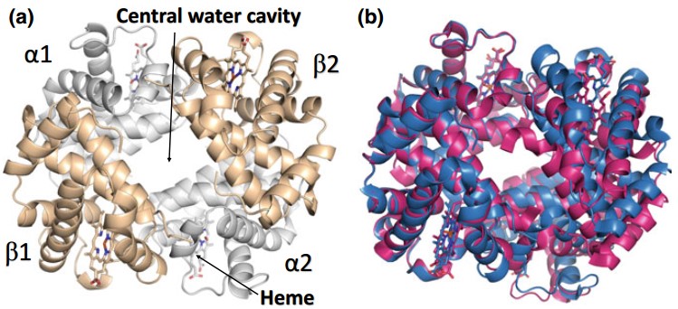 Figure 1. Structural and conformational changes in hemoglobin
(Source: Ahmed MH, et al. 2020)
Humans have three main types of hemoglobin, including adult hemoglobin (HbA), fetal hemoglobin (HbF) and abnormal hemoglobin. HbA is the predominant type of hemoglobin in adults, while abnormal hemoglobin, such as sickle cell hemoglobin (HbS), have structural abnormalities that result in altered red blood cell morphology, which may lead to diseases such as sickle cell anemia. HbF consists of two α-chains and two γ-chains and has a stronger affinity than HbA, allowing the fetus to efficiently obtain oxygen from the mother.
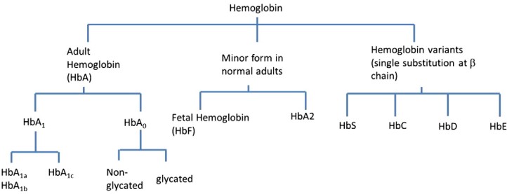 Figure 2. Hemoglobin and its variants
(Source: Ang SH, et al. 2015)
During embryo-to-adult development, ζ, α1 and α2-globin proteins are located in the β-globin chain on chromosome 11, respectively. The ε- and γ-chains form Hb Grower Ⅰ (ζ2ε2), Hb Grower Ⅱ (α2ε2), and Hb Portland (ζ2γ2) hemoglobins in the yolk sac. Gene expression on the β-globin cluster undergoes two very important globin transitions to satisfy the body's need for oxygen supply during development. The first gene expression transition occurs around the 6th week of pregnancy, when embryonic hemoglobin is restricted to the yolk sac stage of development, while γ-globin expression begins to be initiated. At this time HbF consists of 2 α-subunits and 2 γ-subunits and is mainly synthesized in the spleen and liver of the fetus. There is a gradual increase in γ-globin expression from after the 12th week of pregnancy until the birth of the fetus, when HbF levels reach up to 90%. Because of the higher affinity of HbF to bind oxygen, the fetus is able to receive sufficient oxygen from the mother through the umbilical cord even in a less oxygenated environment. After birth, a second globin transition is completed in the body, and γ-globin expression begins to decline and eventually shuts down, at which point β-globin expression begins to increase. At this stage, HbA completes its synthesis as α2β2 in the bone marrow and begins to replace HbF as the main component that makes up human hemoglobin.
 Figure 3. The fetal-to-adult hemoglobin switch
(Source: Sankaran VG, et al. 2013)
Hemoglobin oxygen affinity is the ability of Hb to bind oxygen, which is one of the important characteristics of Hb and is mainly obtained by reading the oxygen dissociation curve. The oxygen dissociation curve is a curve that represents the relationship between Hb oxygen saturation and PO2, and the synergistic effect of Hb gives the oxygen dissociation curve an S-shape. In contrast, monomers such as myoglobin do not have a synergistic effect and therefore have a parabolic oxygen dissociation curve. The Hb oxygen affinity value was defined as the partial pressure of oxygen (P50) corresponding to an Hb oxygen saturation of 50% in the oxygen dissociation curve. Changes in P50 affect the body's oxygen uptake and oxygen acquisition by tissue cells, with normal adults having a P50 of 24-28 mmHg. Elevated P50 represents decreased Hb oxygen affinity and easy release of O2, and decreased P50 represents increased Hb oxygen affinity and easy binding of O2.
The affinity of hemoglobin is regulated not only by oxygen concentration but also by pH and carbon dioxide concentration. When the CO2 concentration increases or the pH decreases, the affinity of hemoglobin for oxygen decreases and the oxygen dissociation curve shifts right. In contrast, when the CO2 concentration decreases or the pH increases, the oxygen affinity of Hb increases and the oxygen dissociation curve shifts left. This phenomenon is known as the Bohr effect. This mechanism ensures that in metabolically active tissues, more oxygen can be released to meet the cellular demand for oxygen.
Changes in temperature also cause changes in Hb oxygen affinity. When the temperature increases, the Hb oxygen affinity decreases, the oxygen dissociation curve shifts to the right, and the P50 increases. When the temperature decreases, the Hb oxygen affinity increases, the oxygen dissociation curve shifts left, and the P50 decreases. The effect of temperature on the oxygen affinity of Hb may be related to the change in the activity of H+, which increases at higher temperatures and reduces the affinity of Hb for O2.
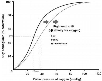 Figure 4. Oxy-hemoglobin dissociation curve
(Source: Min K, et al. 2020)
Increased Hb can be divided into physiological and pathological. Physiological increase is seen in living in the plateau area with low oxygen content for a long time, and concentration of body fluids caused by high temperature and excessive sweating. At high altitude, as the concentration of oxygen in the air decreases, the body develops adaptive responses, including increased erythropoiesis to improve oxygen carrying capacity. This process involves the secretion of erythropoietin (EPO), which promotes erythropoiesis in the bone marrow. Pathological increases are seen in relative increases due to vomiting, high fever, diarrhea and extensive burns, as well as in primary increases such as true erythrocytosis, and secondary increases caused by chronic cardiopulmonary disease, anomalous hemoglobinopathies, and hyperadrenocorticism resulting in hypoxia that leads to a high secretion of EPO.
Several studies have found that increased Hb is associated with hypertension. On the one hand, sustained elevation of blood pressure contributes to the proliferation of vascular elastin fibres and endothelial fibrous tissue. Hypoxia occurs when the vascular lumen narrows. Hypoxia is followed by stimulation of renal secretion of EPO, which increases erythropoiesis and Hb. On the other hand, increased Hb increases blood viscosity and peripheral resistance, which in turn increases blood pressure. Free Hb also binds to nitric oxide and inactivates it, thus preventing vasodilation leading to increased blood pressure.
Glycosylated hemoglobin (HbA1c) is a hemoglobin that binds glucose at the amino- terminus of the β-chain, and plays an important role in glycemic control and risk assessment of macrovascular and microvascular complications as an indicator of average blood glucose concentration. HbA1c is structurally stable and its level changes continuously throughout the life cycle of red blood cells, reflecting the weighted average blood glucose level over the last 2-3 months. The results of a number of current studies have demonstrated that HbA1c is equal to or better than fasting plasma glucose (FPG), and random plasma glucose (RPG) in predicting improvement in cardiovascular disease risk. In a prospective observational study, each 1% reduction in HbA1c among patients with type 2 diabetes was associated with a 14% reduction in the relative risk of myocardial infarction and a 37% reduction in the relative risk of microvascular complications. Another study found that pre-diabetes defined on the basis of HbA1c differentiated the risk of chronic kidney disease, cardiovascular disease, peripheral arterial disease and all-cause mortality better than the FPG-based definition.
Hemoglobin-based oxygen carriers (HBOCs) are a kind of erythrocyte substitutes, which are soluble nanoscale oxygen carriers with oxygen-carrying and releasing functions prepared from matrix-free hemoglobin through chemical modification, etc., and they can replace erythrocytes to deliver oxygen to organs and tissues in the body, and they have the characteristics of no risk of blood type and virus transmission, long storage time and instant availability. The oxygen carrier can replace red blood cells to deliver oxygen to various organs and tissues in the body. HBOCs have the property of rapid oxygen release, and their oxygen release rates are all higher than those of erythrocytes. Based on this property, HBOCs can be used as therapeutic agents in several clinical areas, such as blood transfusion, organ protection, and cerebral ischemia.
References
| Target | Cat. No. | Product Name | Size | Species | Application | Detection Sample | |
| Hemoglobin | DEIA-BJ175 | Human HBμ(Hemoglobin, Mu) ELISA Kit | 96T | Human | Quantitative | Serum, plasma, tissue homogenates, other biological fluids | Inquiry |
| DEIA-BJ127 | Human Glycated hemoglobin A1c ELISA kit | 96T | Quantitative | Serum, plasma, cell culture supernatants, body fluid and tissue homogenate | Inquiry | ||
| DEIABL387 | Cat Hemoglobin ELISA Kit | 96T | Quantitative | Plasma, Serum | Inquiry | ||
| DEIABL394 | Dog Hemoglobin ELISA Kit | 96T | Quantitative | Plasma, Serum | Inquiry | ||
| DEIA686 | Human Hemoglobin ELISA Kit | 96T | Human | Quantitative | Blood, serum, plasma, CSF, cell culture media | Inquiry | |
| DEIA687 | Human Fetal Hemoglobin ELISA Set | 1000T | Human | Quantitative | Serum | Inquiry | |
| HbA1c | DEIA-BJ2475 | Mouse HbA1c(Glycosylated hemoglobin A1c) ELISA Kit | 96T | Mouse | Quantitative | Serum, plasma, tissue homogenates, other biological fluids | Inquiry |
| HBA1 | DEIA3509 | Human HbA1c(Glycosylated hemoglobin A1c) ELISA Kit | 96T | Human | Quantitative | Serum, plasma, tissue homogenates and other biological fluids. | Inquiry |
| HBB | DEIA049J | Hemoglobin (1-point-calibration) ELISA Kit | 96T | Human | Quantitative | Stool | Inquiry |
| HBB/HP | DEIA050J | Hemoglobin/Haptoglobin-Complex (1-point calibration) ELISA Kit | 96T | Quantitative | Stool | Inquiry |
| Target | Cat. No. | Product Name | Expression System | Tag/Conjugate | Application | |
| Hemoglobin | DAG-WT2535 | Hemoglobin (Hb) control | N/A | Unconjugated | Immunoassays | Inquiry |
| Hb | DAG109 | Human Hemoglobin | N/A | Unconjugated | N/A | Inquiry |
| Serum | DAGA-635 | Horse hemoglobin reference serum | Horse | Unconjugated | N/A | Inquiry |
| HbA1c | DAG-WT112 | Hemoglobin A1c Antigen-Low (HbA1c Ag-L) | N/A | Unconjugated | Control/Calibrators | Inquiry |
| DAGA38100 | Human Hemoglobin A1c (HbA1c) | Human Erythrocytes | N/A | Immunoassays | Inquiry |
Loading ......
