Vibrio cholerae is a Gram-negative bacterium belonging to the family Vibrionaceae. Ecological environments are found in coastal waters and estuaries, often associated with zooplankton and shellfish. Currently, more than 200 species of Vibrio cholerae have been identified based on variations in the "O" antigen, and most strains of Vibrio cholerae are not pathogenic. So far, only strains belonging to serogroups O1 and O139 have been associated with epidemic cholera. People become infected by ingesting water or food contaminated with Vibrio cholerae. Most of the Vibrio cholerae ingested is killed by stomach acid. The surviving bacteria colonize in the small intestine and produce cholera toxin, causing people to suffer from acute watery diarrheal disease symptoms. Although water quality, sanitation and clinical treatment of cholera have improved, the disease still kills approximately 100,000 people worldwide each year. An analysis shows that in cholera-endemic countries, approximately 29 million cases and 95,000 deaths occur each year, of which Africa accounts for 60% and 68% respectively.
Currently, the methods used to detect Vibrio cholerae mainly include microbiological methods, molecular biology methods and immunological methods. Compared with microbiological methods, molecular biology methods are more and more widely used, and the combination of these two methods provides a powerful tool for isolation, detection and identification. Immunological methods are more suitable for early detection of infection. For example, during the cholera epidemic in Bangladesh, immunological methods were used to identify V. cholerae in environmental samples when the bacteria in the samples could not be cultured. Currently, although microbiology and molecular biology methods are sufficiently sensitive and accurate, the need for expensive equipment and specialized personnel is an obstacle to their promotion in field and entry-level laboratories. Immunochromatographic assays using colloidal gold nanoparticles as biolabels are a common rapid detection method; however, evaluation of their performance has revealed shortcomings in specificity. Therefore, rapid detection methods that are fast, simple, sensitive and accurate are still needed. In this review, we will introduce internationally recognized routine detection methods and new technologies for V. cholerae detection.
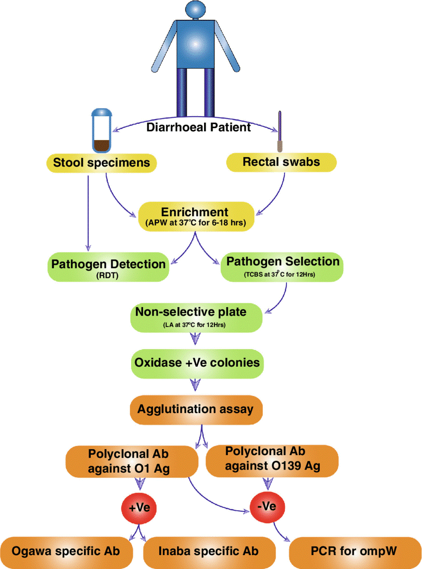 Figure 1. Schematic outline of isolation and identification of V. cholerae from stool specimens.(Source: Ramamurthy T, et al. 2020)
Currently, the gold standard for cholera diagnosis in outbreak management remains isolation of the bacteria from stool samples via selective media followed by biochemical identification and serotyping. Vibrio cholerae does not have high nutritional requirements, can grow well on ordinary culture media, and grows rapidly in alkaline peptone water, so it is often used as a selective enrichment medium, such as TCBS. Glossy colonies with a diameter of 2–4 mm on TCBS can be considered Vibrio cholerae. It takes approximately 8 days to confirm whether a cholera case is present. And these suspicious colonies need to be confirmed by biochemical tests, followed by serogroup confirmation and determination of antibiotic resistance, or by performing PCR using primers developed to target O1 and O139 of genomic DNA to comprehensively evaluate the results. This can determine whether it is Vibrio cholerae.
Microbiological detection methods have good sensitivity and specificity, but their detection time is long, the workload is large, and they are easily affected by the environment, culture conditions and operator subjective factors. They cannot meet the needs of a rapid response system for disease prevention and control, and are not conducive to Rapid clinical diagnosis.
ELISA
Enzyme-linked immunosorbent assay (ELISA) uses antigen-antibody specific binding reactions to detect target samples. Generally, it can be directly detected without separation and is easy to operate. Researchers used the VcNo. 6 antibody to establish a double-antibody sandwich method to detect Vibrio cholerae. The sensitivity of this method reached 103CFU/mL. The antibody specificity test results showed that the specificity of VcNo. 6 was also good. In the matrix addition test, the lowest detection limit is 1 CFU/g sample. Unfortunately, ELISA testing cannot achieve more accurate quantification.
Rapid Detection Technology
The current RDT for Vibrio cholerae is an immunochromatographic test strip detection technology based on gold-labeled antibodies or fluorescent-labeled antibodies. It mainly detects lipopolysaccharides of serogroups O1 and O139 that are prone to causing epidemic cholera. Because it is fast, economical, easy to operate and does not require professional technicians, it can be used as an alternative for cholera diagnosis in environments with poor health and medical conditions.
There is currently a cholera RDT product on the market, CrystalVC, which is a commercial detection kit that uses immunochromatography principles combined with gold-labeled antibodies to detect Vibrio cholerae lipopolysaccharide (LPS) in clinical and environmental samples. Rapid detection of Vibrio cholerae serotypes O1 and O139 directly from stool samples takes only 15–20 minutes. Its detection sensitivity for O1 serogroup is 106CFU/mL and O139 is 107CFU/mL. However, the specificity is low, only about 60%–70%.
The markers used in the above Vibrio cholerae RDT are colloidal gold or fluorescent substances. However, colloidal gold as a marker can only detect optical signals at 10–20 μm on the surface of the nitrocellulose membrane, resulting in a loss of nearly 80%–90% of the signal. In recent years, there have been increasing reports of using magnetic nanomaterials as markers to detect pathogenic microorganisms. Test strips using magnetic nanomaterials as markers detect magnetic signals, and the magnetic analyzer can detect most of the magnetic signals (80%–90%) from the surface to the internal three-dimensional structure of the nitrocellulose membrane. In addition, because biological samples usually have extremely low magnetic background noise, the interference to the detection signal is minimal, and the magnetic signal is very stable and is not interfered by the color of the sample.
Polymerase Chain Reaction (PCR)
In 1987, the invention of PCR enabled highly sensitive detection of bacteria. Because it detects organisms by amplifying the target rather than the signal, it is less prone to false positives. The target DNA can be amplified 1 million times in less than 1 hour, and theoretically the sensitivity can reach the detection of a single target pathogen. Multiplex PCR can detect several pathogenic bacteria at the same time by using different primers to amplify the DNA region encoding the specific genes of each bacterial strain. For example, the virulence genes ctxA and tcp of Vibrio cholerae and other genes such as rfb, ompW, and hly can not only achieve the purpose of efficiently detecting Vibrio cholerae, but also change the false positive phenomenon caused by the insufficient specificity of virulence genes.
Gene Chip
Gene chips are also called "biochips" and can be divided into protein chips, carbohydrate chips and gene chips according to the type of molecular probes fixed on the substrate. In particular, gene chips have become an important analytical tool for current biological detection. They are all based on the specific hybridization between nucleic acid probes and their complementary targets to form stable duplexes or triplexes. Gene chips, consisting of thousands of functionalized probes immobilized on a solid matrix, are comprehensive analytical devices that have been introduced into many fields such as biochemistry and medical diagnosis and can provide accurate, high-throughput results compared to PCR.
Loop-Mediated Isothermal Amplification Technology (LAMP)
LAMP is another variation of nucleic acid-based detection methods. A unique feature of this method is that amplification is performed under isothermal conditions at 60–65°C. LAMP-based assays have been used to detect Vibrio parahaemolyticus, Vibrio vulnificus, Salmonella, and Listeria monocytogenes in seafood and environmental samples.
Biological Sensor
A biosensor is an analytical device that converts biological reactions into electrical signals. It consists of 2 main parts: the bioreceptor or biometric element is used to identify the target object, and the transducer is used to convert the target object into a measurable electrical signal. Biological receptors can be tissues, organelles, microorganisms, etc., and can be transduced through one or a combination of optical, electrochemical, micromechanical, and other technologies. Regarding V. cholerae detection, the biosensors developed so far are mainly electrochemical and optical.
Reference