
Inquiry Basket
 Loading ......
Restriction factors are multiple innate immune proteins expressed in mammalian cells that protect against invading retroviruses, such as HIV-1. Two members of the tripartite motif protein family-TRIM5α and TRIMCyp were identified in 2004 as restriction factors that recognize and intercept incoming retroviruses by binding to the capsid that surrounds and protects the viral core. Here, we summarize how TRIM5 proteins recognize incoming retroviral cores and inhibit virus replication.
As members of the TRIM protein family, TRIM5 proteins contain an amino-terminal (N-terminal) RBCC or tripartite motif, which consists of RING, B-box 2 and coiled-coil domains. This motif is followed by a carboxy-terminal (C-terminal) domain that is required for capsid recognition: SPRY (also known as PRY-SPRY or B30.2) in TRIM5α and cyclophilin A (CypA) in TRIMCyp. The fundamental oligomeric state of TRIM5 is a dimer made up of two coiled-coil domains packed in an antiparallel manner to form an elongated rod that is capped at each end by a B-box 2 domain. The B-box 2 domain also independently mediates self-association, having a plastic oligomerization interface that can form both dimers and trimers. The combined oligomerization properties of the coiled-coil and B-box 2 domains confer on TRIM5 the propensity for higher-order assembly. In vitro, purified TRIM5 protein dimers spontaneously assemble into a hexagonal lattice35. In cells, TRIM5 forms cytoplasmic assemblages or ‘bodies’ when overexpressed, and these are reasonably believed to be made up of the same hexagonal lattice observed in vitro.
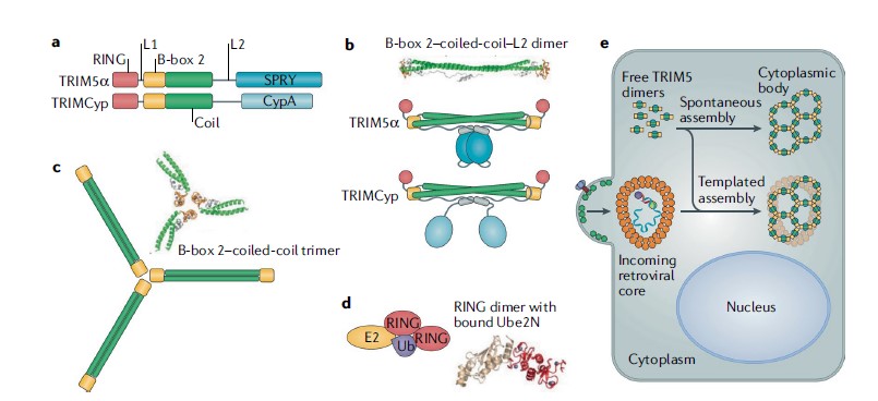 Fig 1. Structural and functional properties of TRIM5
(Source: Nat Rev Microbiol. July 2019)
All retroviral capsids are organized as fullerene structures with hexagonal lattice symmetry, and it is this shared architecture that underlies the mechanism of capsid recognition by TRIM5. Capsid recognition is described by a model in which the SPRY and CypA domains each bind to some epitope on CA.
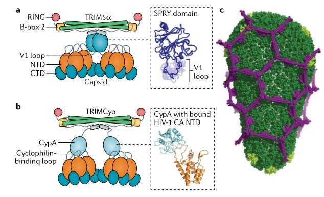 Fig 2. Mechanism of core recognition by TRIM5
(Source: Nat Rev Microbiol. July 2019)
Avid capsid recognition by TRΙM5α Early studies established that TRIM5α does not bind the isolated CA subunit, and that binding can be detected in vitro only when the CA proteins are assembled as capsid-like tubes, other studies showed that the self-association properties of the TRIM5α coiled-coil and B-box 2 domains are critical for restriction activity. In addition, the biochemical analysis of TRIM5α proteins found those proteins assemble spontaneously into hexagonal lattices in vitro. This finding led to the realization that TRIM5α can match both the symmetry and the spacing of the capsid lattice and generate powerful avidity effects. An important unanswered question is precisely how the SPRY domain contacts the CA subunits, and several related studies suggest that the SPRY domain has a β-sandwich fold that displays all four loops on one side, where they can act independently or in concert to bind CA in multiple ways.
Capsid recognition by TRIMCyp The capsid-binding domain of TRIMCyp is homologous to the cellular pep-tidylprolyl isomerase CypA. In HIV-1, CypA binds to the cyclophilin-binding loop that is prominently exposed on the NTD of HIV-1 CA. A key proline residue (Pro90) in this loop binds to the enzyme’s active site. When bound to the assembled CA lattice, CypA has a secondary contact with another NTD in the adjacent CA hexamer. meaning that the TRIMCyp dimer can potentially contact at least four CA subunits simultaneously. Unlike the SPRY domains, the two CypA domains are more flexibly tethered to the coiled-coil domain, and so each TRIMCyp dimer presumably has a wider reach than a SPRY dimer and can sample a larger area to optimize local binding. TRIMCyp might have dispensed with the need for higher-order assembly to efficiently recognize retroviral capsids, consistent with the significantly higher affinity of the CypA domain for the CA subunit.
The viral core is recognized through multivalent TRIM5 binding, which might directly inactivate the virus. Effector functions of the RING domain increase the efficiency of inactivation and recruit cellular degradation machinery that clears the debris. Next, ubiquitylation-dependent pathways activate innate immune pathways to signal the presence of an invader. Restriction by capsid binding. The notion that core stability is optimized to promote reverse transcription and infectivity implies that capsid-binding molecules that perturb this delicate balance can be inhibitory in certain contexts. A bivalent or trivalent capsid-binding protein is intrinsically inhibitory, perhaps by interfering with capsid stability or with critical post-entry interactions of the core.
Binding-induced non-productive uncoating. The idea that uncoating is spatially and temporally coupled with reverse transcription also implies that premature dis-sociation of the core can have negative consequences for the virus. a gradient centrifugation protocol which revealed that yields of pelletable and presumed core-associated HIV-1 CA are significantly reduced in cells that express TRIM5. Loss of particulate CA is accompanied by a corresponding increase in the level of soluble CA, which is taken to indicate disassembly, the loss of reverse transcripts caused by TRIM5 directly correlates with accelerated core dissociation. Some studies suggest that TRIM5 binding TRIM5 binding directly destabilizes the capsid lattice, while others suggest that TRIM5 binding does not intrinsically induce dissociation of the CA lattice, but actually induces global rigidification of the CA lattice, while at the same time inducing structural and dynamic changes that map to large portions of the CA tertiary structure. Therefore, further research is needed in this area.
TRIM5 cytoplasmic bodies (both in the presence and in the absence of viral cores) are linked with ubiquitylation and ubiquitin-dependent cellular degradation machinery such as proteasomes. In the context of restriction, in the presence of proteasome inhibitors, the levels of particulate CA and core components resemble those observed under non-restricting conditions. TRIM5α undergoes self-ubiquitylation, and inhibiting this modification abrogates accelerated uncoating and restores reverse transcription. TRIM5 and HIV-1 cores initially form stable complexes, and that the ubiquitin–proteasome system is required to non-productively accelerate disassembly of the capsid coat. The ubiquitin–proteasome system is not required to inhibit virus replication, and even under conditions where reverse transcription is restored by proteasome inhibitors or RING domain mutations, infectivity is not restored. Thus, either TRIM5 imposes sequential blocks to the post-entry pathway or TRIM5 binding diverts the core into an ‘off-pathway’ trajectory that can-not be corrected by inhibiting the proteasome or disrupting TRIM5 self-ubiquitylation.
TRIM5 is a selective autophagy receptor that directs degradation of the retroviral core through a mechanism termed ‘virophagy’. TRIM5α binds and activates key autophagy effectors. TRIM5 also directly binds LC3 and other Atg8 homologues, which are ubiquitin-like proteins that are conjugated to phosphatidylethanolamine and are well-established markers of autophagosomal membranes. However, autophagy is not required for TRIM5-mediated inhibition of virus infectivity, which parallels observations on the proteasome. It might be that the proteasome and autophagy pathways are redundant or cell type-dependent mechanisms of virus clearance that operate after the core has already been disabled.
| Target | Cat. No. | Product Name | Host | Isotype | Application | |
| TRIM5 | DPABH-26148 | Anti-TRIM5 (aa 50-150) polyclonal antibody | Rabbit | IgG | WB, IHC-P | Inquiry |
| DPAB-DC3565 | Anti-TRIM5 (aa 309-418) polyclonal antibody | Mouse | WB, ELISA | Inquiry | ||
| CABT-B11679 | Mouse anti-Human TRIM5 monoclonal antibody, clone 4C22I3 | Mouse | IgG1 | WB, IHC | Inquiry | |
| CABT-B11680 | Mouse anti-Human TRIM5 monoclonal antibody, clone 3B7 | Mouse | IgG2a | ELISA | Inquiry |
Loading ......
