
Inquiry Basket
 Loading ......
Thymoma is the most common primary tumor in the anterior superior mediastinum, accounting for 20% -40% of all mediastinal tumors in adults. It originates from thymic epithelium, but does not include tumors originating from germ cells, lymphocytes, neuroendocrine cells and adipose tissue. Most thymomas is located in the anterior mediastinum, attached to the pericardium, and a few occur outside the mediastinum, such as pleura, diaphragmatic angle, lung parenchyma, hilar or neck. In 1999, who developed a classification of thymic epithelial tumors. According to the morphology of epithelial cells and the ratio of lymphocytes to epithelial cells, thymoma was classified into a, AB, B1, B2, B3 and C types. Type C is thymic carcinoma, which has obvious cell heteromorphism and loses the special structure of thymus, which is similar to the carcinoma of other organs.
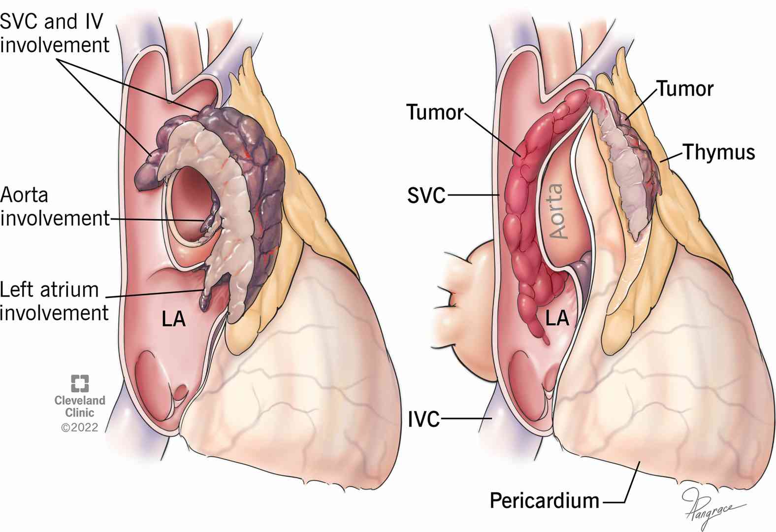 Figure 1. Thymoma.
Thymoma is the most common primary tumor of the anterior superior mediastinum, accounting for 20% -40% of all mediastinal tumors in adults. It originates from thymic epithelium, but does not include tumors originating from germ cells, lymphocytes, neuroendocrine cells and adipose tissue. A few occur outside the mediastinum, such as pleura, diaphragm angle, lung parenchyma, hilar or neck. Thymoma grows slowly, mostly benign, with intact capsule, but it is potentially invasive and easy to infiltrate surrounding tissues and organs. Thymoma is closely related to autoimmune disorders, often accompanied by paraneoplastic syndromes such as myasthenia gravis (mg), various granulocytopenia, red blood cell dysplasia, hypogammaglobulinemia, collagen angiopathy, etc. Foreign literature shows that the annual incidence of thymoma in the population is 0.15/100000, the male to female ratio is 1:1, and the peak age of incidence is 40~50 years old. The incidence of thymoma associated with myasthenia gravis is about 10%~46%, mostly in 30~40 years old. Thymoma in children is rare, but the degree of malignancy is higher. The pathogenesis of thymoma is still unclear. Some scholars believe that the patient's previous history of radiotherapy and EB virus infection may be related to thymoma.
The clinical symptoms of thymoma arise from the compression of the surrounding organs and the unique symptoms of the tumor itself - combined syndrome. Small thymomas are mostly asymptomatic and not easy to be found. When the tumor grows to a certain volume, the common symptoms are chest pain, chest tightness, cough and anterior chest discomfort. The symptoms lasted for a long time. Some patients underwent X-ray examination or some patients found mediastinal tumor shadow during chest X-ray examination or chest radiography. Thymoma which is neglected to be diagnosed often grows to a considerable volume, oppresses the innominate vein or has the appearance of superior vena cava obstruction syndrome. Severe chest pain, rapid exacerbation of symptoms in the short term, severe irritating cough, dyspnea caused by pleural effusion, palpitation and shortness of breath caused by pericardial effusion, and pain in joints and bones all suggest the possibility of thymoma.
Paraneoplastic Syndrome Associated with Thymoma
| Symptoms | Descriptions |
| Neuromuscular syndrome | Myasthenia gravis, eaten Lambert syndrome, myotonic dystrophy, borderline encephalopathy, stiff person syndrome. |
| Gastrointestinal diseases | Chronic ulcerative colitis and localized enteritis |
| Collagen and autoimmune diseases | Systemic lupus erythematosus, sarcoidosis, rheumatoid arthritis, polymyositis, dermatomyositis, pericarditis, Sjogren's syndrome, Raynaud's disease, thyroiditis |
| Skin diseases | Herpes simplex, alopecia, chronic Candida infection |
| Endocrine system diseases | Cushing syndrome, hypothyroidism, Addison disease, hypertrophic osteoarthropathy |
| Diseases of urinary system | Nephropathy, minimal change nephropathy |
| Hematopoietic system diseases | Red cell aplastic anemia, red cell hypoplasia, pernicious anemia, polycythemia, agranulocytosis, multiple myeloma, hemolytic anemia, acute leukemia, t-lymphocytosis |
| Immunodeficiency syndrome | Hypogammaglobulinemia, T-lymphocyte deficiency |
| Symptoms | Descriptions |
| Myasthenia gravis | It has long been found that myasthenia gravis is related to thymus (or thymoma). Myasthenia gravis can be divided into three types, such as blepharoptosis, long-term visual fatigue, diplopia and so on; The trunk type is that the upper limb can not be extended for a long time, and it needs to sit down and rest when walking a little far away; Chewing and swallowing is laborious, and even respiratory muscle paralysis is medullary type. Clinically, the most dangerous is myasthenia crisis. Patients with respiratory muscle paralysis must be assisted by artificial respiration. |
| Simple red cell aplastic anemia | One of the coexisting diseases with thymoma is simple red cell aplastic anemia. Pure red aplastic anemia can be primary, and the cause is unclear. It can also be secondary to drugs, infections and tumors. |
| Nephrotic syndrome nephritis | The relationship between nephrotic syndrome nephritis and thymoma is still unknown. Nephrotic syndrome can be part of the systemic manifestations of certain tumors, such as Hodgkin's disease. The possible explanation is that thymoma reacts with the antigen antibody complex of glomerulonephritis. |
1. Treatment Principle
Thymoma should be surgically removed as soon as it is diagnosed. Thymoma should be removed as soon as possible. Pathological biopsy can be used to guide postoperative treatment for resected thymoma, and postoperative radiotherapy for partial resection can relieve symptoms and prolong the survival time of patients.
2. Matters Needing Attention During Operation
For isolated benign thymoma without adhesion, complete removal is not difficult, and the operation can be successfully completed, but the difficulty should be fully estimated in some complex cases. Thymoma should be explored first to find out the relationship between tumor and adjacent organs before dissection. Thymoma is located at the bottom of mediastinum and the junction of heart and great vessels; Thymoma adhered and infiltrated to the periphery; When the tumor grows, the adjacent tissues and organs are pushed, and the normal anatomical relationship changes; Adhesion and thickening of fibrous connective tissue make it difficult to distinguish from blood vessels, which can cause intraoperative accidental injury to blood vessels and cause massive bleeding.
Early diagnosis and treatment are the key to the prevention and treatment of this disease. At the same time, attention should be paid to prevent the occurrence of various complications. Once they occur, they should be actively treated to prevent the further development of the disease.
Reference
| Cat. No. | Product Name | Host | Isotype | Application | |
| DPAB-DC1666 | Anti-IFI27 (full length) polyclonal antibody | Mouse | WB, ELISA | Inquiry | |
| DPAB-DC2887 | Anti-DAP3 (aa 44-57) polyclonal antibody | Rabbit | IgG | WB | Inquiry |
| DPABH-22858 | Anti-SLC4A3 polyclonal antibody | Rabbit | IgG | WB, IHC-P | Inquiry |
| Cat. No. | Product Name | Size | Species Reactivity | Application | Detection Method | |
| DEIABL443 | anti-Titin-Antibody ELISA Kit | 96T | Human | Qualitative | Inquiry |
| Cat. No. | Product Name | Size | Target | Species | |
| CDBP5044 | AKT1 blocking peptide | 0.05 mg | / | / | Inquiry |
| CDBP0365 | Human AKT3 blocking peptide | 100 g | AKT3 | Human | Inquiry |
| CDBP0364 | Human AKT1 blocking peptide | 50 g | AKT | Human | Inquiry |
| DAG-KO002 | AKT1 Knockout Cell Lysate | 100 g | / | / | Inquiry |
| CDBP5044 | AKT1 blocking peptide | 0.05 mg | / | / | Inquiry |
Loading ......
