
Inquiry Basket
 Loading ......
Fibroblast Growth Factor 1 (FGF1), also known as aFGF, is an early identified member of the FGF family. FGF1 is a critical regulator of physiological and pathological processes. FGF1 is a strong signaling molecule that can bind to cell-surface Fibroblast Growth Factor Receptors (FGFRs) and Heparan Sulfate Proteoglycans (HSPGs) to initiate complex downstream signaling cascades that govern cell proliferation, differentiation, migration and survival.
Browse all FGF1 related products
FGF family is a large group of polypeptide signalling molecules with key regulatory functions in the embryonic development, tissue homeostasis, repair upon injury and the pathophysiology of diverse diseases in vertebrates. FGF1 is among the first identified and characterised FGFs and as a result has been the focus of extensive interest due to its broad spectrum of biological activities. FGF1 is unusual among the FGFs in lacking a classical signal peptide sequence and release is independent of the canonical endoplasmic reticulum-Golgi secretory pathway.
FGF1 is a relatively small protein of approximately 17-18 kDa, with 155 amino acids. It adopts the classic β-trefoil fold structure, composed of 12 antiparallel β-strands, that form a very stable and compact barrel-like core. The core is thought to be necessary to ensure stability in the extracellular space from protease degradation and thermal denaturation. The molecular surface is enriched with two main functional sites, the receptor-binding site and the heparin-binding site. The receptor-binding site has the capability of high-affinity binding to the specific FGFRs, the first step in the initiation of intracellular signaling. The heparin-binding site is a sequence enriched with positively charged amino acids, that interacts with the negatively charged HS chains of the cell surface and extracellular matrix. Heparin-binding activity is necessary for stabilizing its conformation and a prerequisite for receptor activation. HS-binding protects FGF1 from protease degradation, increases its half-life, and is also necessary for its concentration gradient formation in tissue.
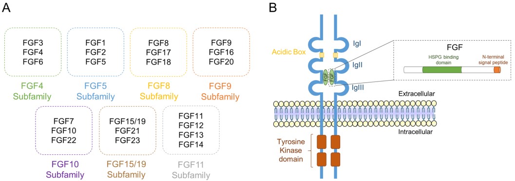 Figure 1. Fibroblast growth factor (FGF) phylogeny and receptor structure
(Source: Tomé D, et al. 2023)
FGF1 was among the first highly potent mitogens to be identified. Mitogens are simply molecules that can cause cells to undergo cell division. FGF1 can induce proliferation of almost any mesoderm and neuroectoderm-derived cells including fibroblasts, vascular endothelial cells, smooth muscle cells, chondrocytes and neurons. FGF1 promotes cell cycle G1 to S phase transition via classical RAS-MAPK signal transduction pathway, thus inducing cell division. Meanwhile, via the PI3K-AKT signaling pathway, FGF1 also potently inhibits apoptosis to allow cell survival in even harsh environments. In some cells and environments, FGF1 can also promote cell differentiation, for example, induce differentiation of neural stem cells into neurons, and help normal tissue and organ development. FGF1 has been found to be one of the most potent pro-angiogenic factors identified. FGF1 directly targets vascular endothelial cells to induce proliferation, migration, and tubulogenesis. FGF1 can also upregulate other critical pro-angiogenic factors such as Vascular Endothelial Growth Factor (VEGF) and act in a synergistic fashion with these proteins to promote angiogenesis. This can be beneficial in wound healing and in treatment of ischemic diseases, but is often a target in cancer research as a mechanism of tumor growth and metastasis.
Following tissue injury, the release of FGF1 is a critical signal initiating the repair process. It further induces angiogenesis to re-establish the blood supply, as well as attract and activate fibroblasts that produce extracellular matrix to fill the tissue defect. In particular, FGF1 has a significant neuroprotective and regenerative effect in nervous system injuries. FGF1 has been shown to protect neurons from diverse insults such as excitotoxicity, oxidative stress, and ischemia and to promote axonal growth and neural circuitry reconstruction.
FGF1 has more recently become a broadly studied metabolic regulator. FGF1 was shown to cause sustained reductions in circulating glucose and improvements in insulin resistance in diabetic mice after a single peripheral administration, and some of the efficacy endpoints were superior to those of some currently used glucose-lowering therapies. To achieve its systemic effects on glucose and lipid metabolism, FGF1 acts on the adipose tissue, the liver, and the central nervous system to stimulate adipose tissue remodeling, inhibit hepatic gluconeogenesis, and increase glucose uptake and disposal in peripheral tissues. The recognition of FGF1 as a key metabolic regulator has created an exciting new avenue for therapeutic intervention in type 2 diabetes and other related metabolic diseases. Its translation into the clinic is hampered by mitogenic side effects.
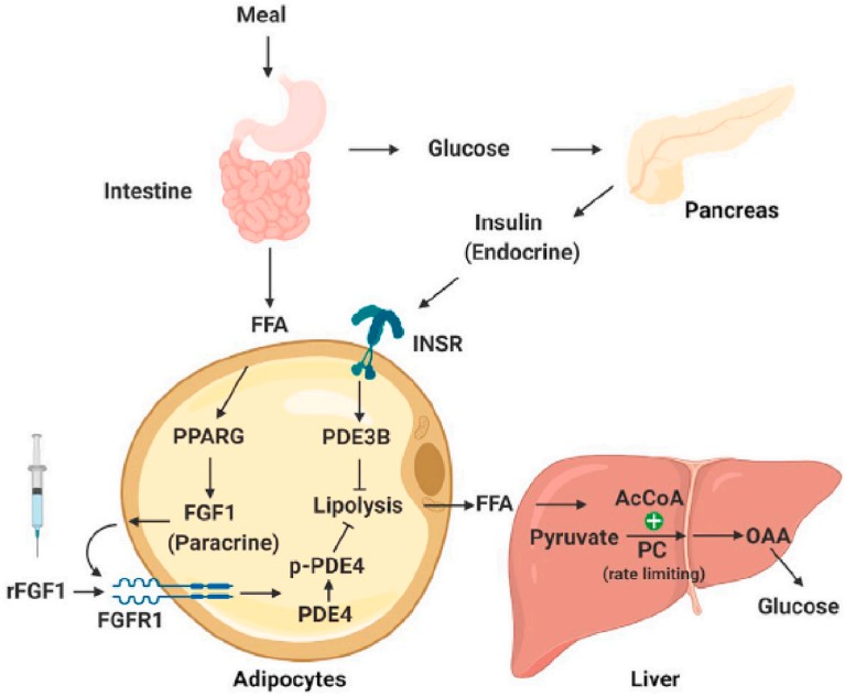 Figure 2. Model of FGF1-induced suppression of lipolysis and HPG.
(Source: Jamal SB, et al. 2024)
The functions of FGF1 are executed by an elaborated hierarchical signal transduction machinery. At its center are FGFRs and co-receptor HSPGs. FGFRs are single-pass transmembrane receptor tyrosine kinases (RTKs), which include FGFR1, FGFR2, FGFR3 and FGFR4. The extracellular domains of FGFRs comprise of 2 or 3 Ig-like domains that mediate binding to FGF ligands. Alternative splicing of the third Ig domain of FGFR1-3 results in two different isoforms, "b" or "c" (beta or gamma), which are responsible for binding specificity to the various FGF ligands. A defining property of FGF1 is that it is a "broad-spectrum" binder, in that it can bind to multiple FGFR isoforms (primarily FGFR1c, FGFR2c, FGFR3c) and FGFR4. At a molecular level this explains the versatile nature of FGF1. In contrast, signal activation by FGF1 is strictly HSPG dependent. Two FGF1, two FGFR and HSPG chains form a stable symmetric dimer complex (2:2:1 or 2:2:2 stoichiometry) during signal activation. HSPGs thus provide the platform or "molecular scaffold" that not only stabilizes the FGF1: FGFR interaction but also determines binding specificity, dictated by unique sulfation patterns.
After the formation of the ternary complex, the two adjacent FGFR molecules dimerize. They trans-phosphorylate and activate their intracellular tyrosine kinase domains. The activated FGFRs then phosphorylate a variety of downstream adaptor proteins, including FRS2α (Fibroblast Growth Factor Receptor Substrate 2α) and PLCγ (Phospholipase Cγ), which initiate several parallel signaling pathways:
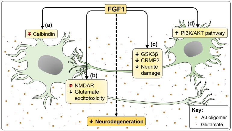 Figure 3. Neuroprotective effects of FGF1 in in vitro AD models.
(Source: Alam R, et al. 2022)
As FGF1/FGFR signaling is so central to cancer, there are numerous FGFR pathway inhibitors that are being developed, and have been a very hot topic in oncology recently. Small molecule tyrosine kinase inhibitors (TKIs) are capable of penetrating the cell membrane and directly binding in a competitive manner to the ATP-binding site of the FGFR kinase domain, and so inhibit kinase activity. Non-selective TKIs (i.e. also inhibiting FGFR and VEGFR, etc.) and highly selective pan-FGFR inhibitors can be differentiated based on selectivity. As a different strategy, monoclonal antibodies act extracellularly. One such approach is directed against FGFRs and inhibits signaling either by blocking the binding sites of FGF1 or by inducing internalization and degradation of the receptors. The other approach is directed against the FGF1 ligand and thus directly neutralizes extracellular FGF1 and prevents it from binding to its receptors. As another strategy, FGF "traps" (soluble FGFR fusion proteins containing the extracellular, ligand binding domain of FGFR) can be used. These "traps" can bind to ligands such as FGF1 with high affinity in the bloodstream and sequester them from binding to functional receptors on the cell surface and thereby inhibit the signaling pathway. Some of these inhibitors have been found to have clinical efficacy in the treatment of different solid tumors with FGFR gene amplification, fusions, or mutations. However, drug resistance is a major issue.
References
| Target | Cat. No. | Product Name | Size | Species | Application | Detection Sample | |
| FGF1 | DEIA-BJ2858 | Canine Acidic fibroblast growth factor 1 ELISA Kit | 96T | Quantitative | Serum, plasma, cell culture supernatants, body fluid and tissue homogenate | Inquiry | |
| DEIA-BJ2358 | Mouse FGF1(Heparin-binding growth factor 1) ELISA Kit | 96T | Mouse | Quantitative | Serum, plasma, tissue homogenates, other biological fluids | Inquiry | |
| DEIA-BJ2752 | Porcine Acidic fibroblast growth factor 1 ELISA Kit | 96T | Quantitative | Serum, plasma, cell culture supernatants, body fluid and tissue homogenate | Inquiry | ||
| DEIA-BJ2041 | Rat Acidic fibroblast growth factor 1 ELISA Kit | 96T | Quantitative | Serum, plasma, cell culture supernatants, body fluid and tissue homogenate | Inquiry | ||
| DEIA3016 | Human FGF acidic ELISA Kit | 96T | Human | Quantitative | Cell culture supernatants, serum, plasma, urine | Inquiry | |
| DEIA5251 | Human bFGF (for Lysates) ELISA kit | 96T | Human | Quantitative | Cell lysates, tissues lysates | Inquiry | |
| DEIA-BJ896 | Human FGF1(Heparin-binding growth factor 1) ELISA Kit | 96T | Human | Quantitative | Serum, plasma, tissue homogenates, other biological fluids | Inquiry |
| Target | Cat. No. | Product Name | Host | Isotype | Application | |
| FGF1 | DCABH-546 | Anti-FGF1 monoclonal antibody, clone 5E3 | Mouse | IgG1 | WB, ICC/IF | Inquiry |
| CABT-B9179 | Mouse anti-Human basic FGF monoclonal antibody, clone 7GHG | Mouse | IgG2a | WB, IF, IHC, IP | Inquiry | |
| DPABH-22954 | Anti-FGF1 (aa 16-155) polyclonal antibody | Rabbit | IgG | WB, IP, ELISA, IHC-P, Neut | Inquiry | |
| CABT-NS1695 | Rabbit Anti-Canine FGF1 Polyclonal Antibody | Rabbit | IgG | ELISA | Inquiry | |
| ABPR-0338 | Human FGF1 ELISA Matched Antibody Pair | ELISA | Inquiry | |||
| DPABH-08295 | Anti-FGF1 (full length) polyclonal antibody | Mouse | IgG | WB, ICC/IF | Inquiry | |
| DMABT-H13741 | Anti-FGF1 monoclonal antibody, clone 3F23 | Mouse | IgG1 | WB, IHC, IF, IP, sELISA, ELISA | Inquiry | |
| DCABH-6606 | Anti-FGF1 monoclonal antibody, clone FQ21031 | Rabbit | IgG | WB, IHC-P | Inquiry | |
| DCABH-6652 | Anti-FGF1 monoclonal antibody, clone LU226 | Rat | IgG2a | WB, ELISA | Inquiry | |
| DCABH-7670 | Anti-FGF1 monoclonal antibody, clone FQS2289 | Rabbit | IgG | WB, IHC-P | Inquiry | |
| DCABH-955 | Anti-FGF1 monoclonal antibody, clone 2G0 | Mouse | IgG1 | WB, IHC-P, ICC/IF | Inquiry |
| Target | Cat. No. | Product Name | Expression System | Tag/Conjugate | Application | |
| FGF1 | DAG-WT3514 | Recombinant Human Fibroblast Growth Factor (FGF1) | E. coli | N/A | N/A | Inquiry |
Loading ......
