
Inquiry Basket
 Loading ......
Researchers named ST2 "Suppression of Tumorigenicity 2" due to its accidental discovery during tumor research. Researchers discovered in 1989 that ST2 gene overexpression decreased cellular tumorigenicity during mouse fibroblast tumor suppression gene screening. Subsequent studies, however, have redefined its core functionality: ST2 which is formally known as Growth Stimulation Expressed Gene 2 demonstrates its vital function in cellular proliferation and tissue repair. ST2's biological functions now link its characteristics to cardiovascular diseases through its effects on inflammatory signaling and extracellular matrix control.
ST2 belongs to the interleukin-1 receptor family known as IL-1R family. Alternative splicing produces several ST2 isoforms with the main types being membrane-bound ST2L (ST2 ligand-binding form), soluble ST2 (sST2), ST2V, and ST2LV. The ST2L form contains a full transmembrane receptor setup which includes an extracellular immunoglobulin-like domain beside a transmembrane section that leads into an intracellular Toll/IL-1 receptor (TIR) domain. ST2L functions to mediate IL-33 signals while being mainly expressed on Th2 lymphocytes, mast cells, and cardiac fibroblasts. In contrast, sST2 lacks transmembrane and intracellular domains due to exon 8 splicing, functioning as a decoy receptor that antagonizes IL-33 signaling through competitive binding. sST2 circulates systemically in blood and extracellular matrices, with studies demonstrating its robust secretion by fibroblasts under myocardial mechanical stress, exhibiting a positive correlation with ventricular remodeling severity. The ST2V isoform, restricted to specific cell types like placental cells, remains poorly characterized functionally.
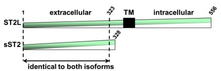 Figure 1. The ST2 gene encodes 2 main protein isoforms
(Source: Pascual-Figal DA, et al. 2015)
Under homeostatic conditions the interaction between ST2L and IL-33 activates MyD88-dependent signaling pathways resulting in NF-κB and MAPK phosphorylation which triggers Th2-type immune responses through IL-4, IL-5, and IL-13 secretion to support tissue repair and regulate inflammation. Tissue restoration during cutaneous wound healing accelerates as fibroblast multiplication is stimulated by the IL-33/ST2L axis. Cardioprotective benefits from this pathway include reduced myocardial hypertrophy and fibrosis. An overabundance of sST2 expression blocks the positive effects by causing collagen buildup alongside ventricular changes.
Cardiac and fibroblast cells emit high quantities of sST2 as a response to mechanical and oxidative stress together with inflammation during cardiovascular disease conditions. sST2 exacerbates disease progression through dual pro-fibrotic mechanisms: sST2 advances disease progression via two pro-fibrotic mechanisms that involve blocking anti-fibrotic IL-33/ST2L signaling and triggering macrophage-derived pro-fibrotic cytokine release. Patients with dilated cardiomyopathy present strong clinical evidence that shows elevated sST2 levels have a significant correlation with the severity of myocardial interstitial fibrosis. Furthermore, coronary artery disease patients demonstrate a linear relationship between sST2 concentrations and ischemic scar burden, establishing its utility as a dynamic biomarker for monitoring ventricular remodeling trajectories.
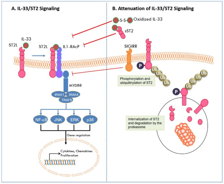 Figure 2. Activation and attenuation of the IL-33/ST2L complex
(Source: Homsak E, et al. 2020)
The biomarker sST2 presents less dependence on kidney function and better biological precision following acute coronary syndrome which positions it as a superior prognostic tool for acute myocardial infarction relative to C-reactive protein and NT-proBNP. The risk of 30-day readmission for patients who suffered a non-ST-segment elevation myocardial infarction (NSTEMI) increases by 3.6 times if their sST2 levels reach 35 ng/mL or higher. Integration with GRACE scores further improves predictive accuracy. The study found that myocardial infarction patients in the high-sST2 group showed significantly higher cardiovascular mortality rates together with more frequent heart failure occurrences, and a 2.5-fold increase in major adverse cardiovascular events (MACE) confirmed sST2 as an independent predictor for one-year cardiovascular death. Research through meta-analyses confirms these results by demonstrating high baseline sST2 levels can forecast short-term and long-term negative results for ACS patients.
sST2 demonstrates utility in identifying myocardial infarction patient subgroups likely to benefit from mineralocorticoid receptor antagonist (MRA) therapy. Eplerenone functions as a selective MRA to reduce post-infarction ventricular remodeling and shows better treatment outcomes in patients who present with high baseline sST2 levels. Optimizing β-blocker doses for ST-segment elevation myocardial infarction patients can be informed by baseline sST2 measurements because increased sST2 levels show better therapeutic outcomes with higher β-blocker dosages. Emerging evidence positions sST2 as a dynamic monitoring biomarker for evaluating management efficacy in both STEMI and heart failure care pathways.
The progression of cardiovascular diseases culminates in heart failure which presents as poor cardiac output along with chronic damage to kidneys, lungs, and liver that significantly reduces patient quality of life. The release of IL-33 during myocardial stretching allows it to bind with transmembrane ST2L which starts cardioprotective signaling that opposes both fibrosis and hypertrophy. Conversely, IL-33 binding to soluble ST2 (sST2)-a decoy receptor-triggers opposing effects, exacerbating fibrotic and hypertrophic pathways. Emerging evidence suggests sST2 may serve as a surrogate marker for pulmonary and vascular congestion in HF.
A study of acute HF (AHF) patients with renal dysfunction revealed an independent inverse correlation between sST2 levels and diuretic response within 24–72 hours. In patients undergoing invasive hemodynamic monitoring-guided therapy, elevated sST2 concentrations predicted impending failure to reduce filling pressures and adverse clinical events. Acute decompensated HF patients with sST2 >35 ng/mL faced a 1-year mortality hazard ratio (HR) of 2.8, while those with persistently high sST2 exhibited a 90-day mortality rate of 42%. Additionally, β-blocker dose titration guided by sST2 levels (e.g., metoprolol escalation from 47.5 mg to 190 mg) reduced rehospitalization rates by 15%, demonstrating its potential for personalized therapeutic optimization.
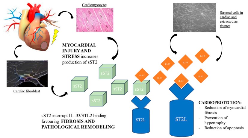 Figure 3. Pathological role of sST2 in promoting fibrosis and ventricular remodeling
(Source: Riccardi M, et al. 2023)
The right ventricle experiences sustained afterload elevation throughout pulmonary arterial hypertension (PAH) development which results in compensatory hypertrophy that leads to eventual right heart failure. Recent studies show that sST2 acts as a new biomarker for right ventricular dysfunction because its concentration changes align closely with hemodynamic decline, clinical results and treatment effects in patients with PAH. Mechanistic studies demonstrate that IL-33 secreted by pulmonary arterial endothelial cells under hypoxic stress acts paracrinely on ST2L receptors expressed on right ventricular cardiomyocytes, activating the PI3K/Akt/mTOR pathway to suppress apoptosis and enhance energy metabolism.
Paradoxically, serum sST2 levels are markedly elevated in PAH. This elevation involves dual mechanisms: 1) Direct mechanical stimulation from right ventricular pressure overload induces sST2 secretion by cardiac fibroblasts, promoting collagen deposition through IL-33/ST2L pathway antagonism; and 2) IL-13 released by M2 macrophages infiltrating remodeled pulmonary vasculature further amplifies sST2 expression, establishing a pro-fibrotic positive feedback loop. Clinical observations reveal that PAH patients with sST2 concentrations exceeding 50 ng/mL exhibit a 3.2-fold increased risk of right ventricular decompensation within one year, underscoring its prognostic utility in risk stratification.
Clinical cohort studies confirm significant correlations between sST2 concentrations and right ventricular functional parameters. Patients with sST2 levels above 50 ng/mL face a 3.2 times higher one-year mortality risk according to the REVEAL 2.0 risk assessment model independent of traditional biomarkers such as NT-proBNP. Notably, sST2 demonstrates superior sensitivity to right ventricle-pulmonary artery uncoupling compared to echocardiographic indices: According to CMR data analysis there exists an inverse correlation between sST2 concentration and right ventricular ejection fraction where each 10 ng/mL rise in sST2 results in a 1.8% decline in right ventricular longitudinal strain. Furthermore, serial sST2 monitoring enables early detection of clinical deterioration. PAH patients showing <15% reduction in sST2 after three months of targeted drug therapy face an 82% probability of inadequate 6-minute walk distance improvement, necessitating therapeutic regimen modification.
Combining multiple markers improves the clinical application of sST2. Synergistic analysis with NT-proBNP reveals unique prognostic insights: The diagnostic model which combines sST2 with NT-proBNP and Galectin-3 (Gal-3) biomarkers shows improved sensitivity and specificity to distinguish between compensated right heart failure and decompensated right heart failure compared to separate biomarkers. Patients show a 3.2 times greater chance of developing diuretic resistance when their sST2/NT-proBNP ratio exceeds 0.25. Patients exceeding both sST2 and NT-proBNP thresholds show 42.1% mortality in one year while those under both limits have 4.2% mortality. The quantitative risk stratification system establishes a reliable foundation for tailoring therapeutic decisions to individual patients.
In ACS, time-series analyses of ST2 and high-sensitivity troponin T (hs-cTnT) reveal critical temporal complementarity. sST2 concentration peaks typically lag hs-cTnT elevations by 12–24 hours, a kinetic disparity offering novel insights into myocardial injury progression. Clinically, dual-marker models improve sensitivity for predicting 30-day major cardiovascular events from 68% (single-marker) to 92%, with combined trajectory analysis reducing myocardial salvage index estimation errors to <8%. This temporal stratification enhances early risk assessment and therapeutic monitoring in ACS management.
Cross-pathway biomarker combinations expand clinical utility. The combined use of sST2 (myocardial fibrosis), hs-CRP (inflammation), and GDF-15 (cellular stress) results in a hazard ratio of 7.3 (95% CI 4.1–13.0) for predicting all-cause mortality among patients with chronic heart failure. A multi-organ injury panel which includes cystatin-C for renal function assessment together with FABP3 for intestinal barrier evaluation successfully forecasts the duration of ICU stays in critically ill patients. Those positive for all three biomarkers exhibit an average ICU duration 4.8 days longer than biomarker-negative counterparts, demonstrating the predictive power of mechanistically diverse biomarker integration.
Emerging advancements demonstrate that machine learning (ML)-enabled multi-biomarker models are overcoming the limitations of conventional threshold-based approaches. An ML algorithm incorporating six biomarkers-sST2, NT-proBNP, GDF-15, and three others-achieves risk stratification accuracy exceeding 90%, reducing prediction errors by 19.3% compared to traditional scoring systems. These innovative strategies not only enhance early warning capabilities for cardiovascular events but also pioneer new pathways toward truly personalized medicine.
References
| Target | Cat. No. | Product Name | Size | Species | Application | Detection Sample | |
| IL1RL1 | DEIA8143 | Human ST2(Syntenin 2) ELISA Kit | 96T | Human | Quantitative | Serum, plasma, tissue homogenates and other biological fluids | Inquiry |
| DEIA2910 | Mouse ST2/IL-1 R4 ELISA Kit | 96T | Mouse | Quantitative | Cell culture supernatants, serum, plasma | Inquiry | |
| ABPR-ZB077 | Human ST2/IL-1 RL1 Antibody Pair Set | 5 Plates, 15 Plates | Human | sELISA | Inquiry |
| Target | Cat. No. | Product Name | Expression System | Tag/Conjugate | Application | |
| ST2s | DAG-WT131 | Recombinant Human ST2s protein [His] | Mammalian cells | His | ELISA; WB; IHC | Inquiry |
| DAG-WT3243 | Recombinant Human ssT2 | CHO cells | His | ELISA | Inquiry | |
| IL1RL1 | DAG-ST2 | Recombinant Human ST2 protein | Mammalian cells | His | ELISA | Inquiry |
| DAG-WT2625 | Growth stimulation expressed gene 2 (ST2) control | N/A | Unconjugated | Immunoassays | Inquiry |
Loading ......
