
Inquiry Basket
 Loading ......
Ricintoxin (RT) is one of the most powerful plant protein toxins extracted from the seeds of the plant Ricinus communis. Eukaryotic animal cells are sensitive to RT, and mg-level doses can cause poisoning and even death in animals and humans. The molecular structure of RT is a heterodimer of glycoprotein, which consists of two subunits connected by disulfide bonds. Among them, the A subunit is the toxic polypeptide chain A (RTA), which has the active site of N-glycosidase that inhibits protein synthesis. It can cut off the eukaryotic cell ribosome 28SrRNA from adenine 4324, causing protein translation to stop and causing cell death; The B subunit is the lectin B chain (RTB), which is a binding chain. RTB binds to glycoproteins or glycolipids on the surface of target cells, mediating the endocytosis of toxins into the endoplasmic reticulum and releasing RTA into the cytoplasm. Studies have shown that RT is very toxic, with an intraperitoneal median lethal dose of about 3.0 μg/kg in mice, and its toxicity is 380 times that of organophosphorus pesticides. Therefore, the development of rapid and effective detection methods is particularly important and has become a research hotspot around the world. Based on the physical and chemical properties, biochemical properties and immunological properties of RT, researchers have developed various analytical methods to detect RT, including immune absorption analysis, biomass spectrometry analysis and biosensor analysis.
The immunolabeling method is a detection method that biologically labels antigens or antibodies and uses the characteristics of RT antigen sites to specifically bind to antibodies. It is the most commonly used technical means to detect RT. Commonly used immune antibody labeling methods include radioimmunoassay, enzyme-linked immunosorbent assay (ELISA), colloidal gold labeling method and immunoPCR method.
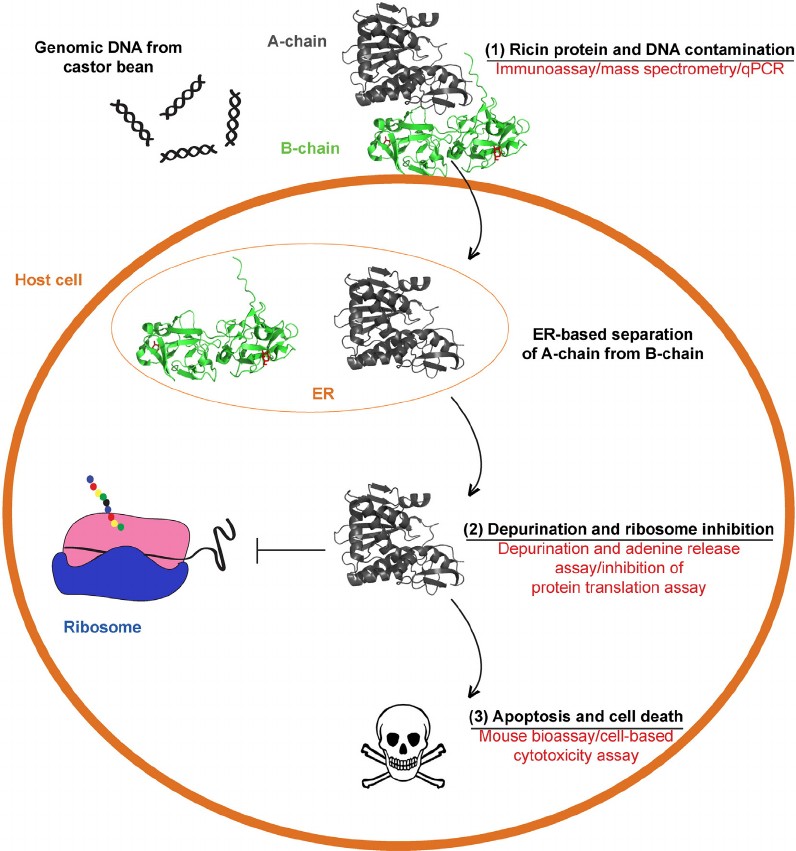 Figure 1. Ricin detection methods in relation to biological steps that occur during ricin intoxication.(Source: Bozza WP, et al. 2015)
Radioimmunoassay (RIA) is an in vitro radionuclide detection method that combines the measurement method of radionuclides with the basic principles of immune response. It is highly sensitive and accurate. Radioimmunoassay is one of the earliest methods to detect low-concentration RT, which uses radioactive I125-labeled RT. Based on rabbit antiserum in buffer, it can be used to quantify RT as low as 100pg. Although radioimmunoassay has good sensitivity, its main disadvantages are the long incubation time, difficulty in dissolving isotopes, and the serious impact of radioactive elements on the environment. These limitations make it less popular compared to ELISA. The radioimmunoassay method can detect RT in the sample on the order of 100pg, and is suitable for detecting RT content in the blood of patients with RT poisoning. However, the disadvantages of this method are that the operation and processing are relatively cumbersome, the equipment requirements are higher than those of the enzyme-linked immunoassay, and the requirements for the professionalism of personnel and the processing of radioactive elements limit the wide application of this method.
With the further development of biomarker technology, researchers have successfully established the ELISA method. The ELISA method is based on antibodies with different antigenic epitopes of RT and detects toxins through the reaction between antigens and antibodies. This method has the characteristics of wide adaptability, easy operation, short detection cycle and high specificity and sensitivity. In addition, the sandwich ELISA method proposed in recent years has further reduced the detection limit.
ELISA is a relatively mature detection method. Experimenters do not need to undergo special training or use special equipment. At the same time, it ensures the speed and accuracy of biological sample poisoning detection, providing technical support for the rapid detection of RT poisoning. Although the assay is less sensitive than reported amplification or chemiluminescent immunoassays, the assay is sufficient to support in vivo RT distribution analysis and on-site RT contamination monitoring.
The colloidal gold labeling method is a new immunolabeling technology that uses colloidal gold as a tracer marker or chromogenic reagent for antigen-antibody reactions. Because it does not have problems such as endogenous enzyme interference and radioactive isotope contamination, and it uses different particles Small and large colloidal gold can also be double or even multiple labeled to make the positioning more accurate, and the colloidal gold biosensor can be used to read the signal and fit the detection curve to complete quantitative detection. In recent years, relevant researchers have tested contaminated samples containing RT, and the detection sensitivity, specificity and stability are good. Some researchers have established an immunochromatographic method for rapid detection of RT. The method is to use rabbit anti-RT polyclonal antibodies as labeled antibodies, label colloidal gold particles, and coat RT antibodies (RCA) on nitrocellulose membranes as detection lines, coated with goat anti-rabbit IgG antibody as a quality control line, and used a colloidal gold biosensor to read the signal, fit the detection curve, and complete quantitative detection. Simulate the addition of RT to common foods to evaluate the detection ability of the method. The results show that the colloidal gold immunochromatography test strip can complete the detection within 15 minutes, with a sensitivity of 0. 925 μg/ml, a linear range of 0. 925~14. 8 μg/ml, and good specificity and stability. Take animal serum, milk, apple juice, orange juice, biscuits, jelly and milk powder to simulate contamination samples respectively, treat them with sample diluent and then add RT. The results show that the test strip can still detect 0.925 μg/ml for the above simulated contaminated RT.
Immuno-PCR (IPCR) is a micro-antigen detection technology based on the highly specific reaction of antigen-antibody binding and the extremely high sensitivity of PCR reaction. IPCR was established by Sano in 1992. This method is essentially an improved ELISA that uses PCR instead of enzyme reaction to amplify and display the antigen-antibody binding rate. Just by introducing PCR, the sensitivity of the existing antigen detection system can be improved by at least several order of magnitude, it is the most sensitive method in RT detection at this stage. IPCR can represent a simple method to detect mature proteins in the immune systems and tissues of animals and humans and can assess the pharmacokinetic behavior of immunotoxins. Comparing the IPCR method with traditional immunological methods, PCR and polymerase chain reaction post-analysis require more time for detection, use more expensive reagents, and increase reagent consumption, making this technology less attractive. However, these limitations are offset by higher sensitivity, enabling a wider range of applications.
Biological mass spectrometry (MS) is becoming the main supporting technology for protein identification and analysis. It performs composition and structure analysis by measuring the mass-to-charge ratio (m/z) of sample ions. The number of protein spots separated by two-dimensional gel electrophoresis is large and the sample amount is small. The identification method is to search the protein database based on various attribute parameters of the protein, such as relative molecular mass, isoelectric point, sequence, amino acid composition and peptide mass fingerprint spectrum, to find proteins that match these parameters.
Biosensors combine biotechnology and electronic technology to convert the chemical signals of biologically sensitive substances generated by the specific combination of the object to be measured and the recognition element into electrical signals, optical signals, etc., so as to achieve the purpose of analysis and detection. This method does not require special markers and has the characteristics of good selectivity, simple operation, and real-time online monitoring.
Reference
| Target | Cat. No. | Product Name | Host | Isotype | Application | |
| Ricin communis | DPATB-H82834 | Anti-Ricin communis polyclonal antibody | IgG | WB, ELISA | Inquiry | |
| Grain Ricin chain A | DPAB2084 | Anti-Grain Ricin chain A polyclonal antibody | IgG | ELISA, WB, Neut | Inquiry | |
| Grain Ricin chain B | DPAB2085 | Anti-Grain Ricin chain B polyclonal antibody | IgG | ELISA, WB, Neut | Inquiry | |
| Ricin | CABT-54812MR | Anti-RCOM_2159910 A Chain monoclonal antibody, clone RA999 | IgG1 | ELISA, WB | Inquiry | |
| DPAB1157 | Anti-Ricin polyclonal antibody | IgG | ELISA | Inquiry | ||
| DPAB4164 | Anti-ricin monoclonal antibody, clone MMH | ELISA, IA, IHC | Inquiry | |||
| Ricin RCA60 | DCAB-TJ081 | Magic™ Anti-Ricinus communis Ricin RCA60 Monoclonal antibody, Clone SC000[Biotin] | IgG1 | EIA, ELISA(Det), WB | Inquiry | |
| DCAB-TJ158 | Magic™ Anti-Ricinus communis Ricin RCA60 Monoclonal antibody, Clone SB000 | IgG1 | EIA, ELISA(Det), WB | Inquiry | ||
| DCAB-TJ159 | Magic™ Anti-Ricinus communis Ricin RCA60 Monoclonal antibody, Clone SC000 | IgG1 | EIA, ELISA(Cap), WB | Inquiry |
Loading ......
