
Inquiry Basket
 Loading ......
Iodination of tyrosine is both biologically relevant and chemically useful post-translational modification (PTM). Iodination of protein tyrosine residues is considered one of the simplest chemical modifications of such complex biomolecules. This type of modification has a rich history and is used for a variety of purposes. Its simplicity and unique properties make it a valuable tool in biological research, aiding in tracking biological samples or cancer treatments, crystallography experiments, and mapping protein surface structures using mass spectrometry.
Protein iodination relies on the incorporation of radioactive iodine isotopes, such as iodine-125 (125I), iodine-131 (131I), or iodine-123 (123I), into proteins through chemical or enzymatic methods. The most commonly used approach is the chloramine-T method, where iodine is introduced using chloramine-T as an oxidizing agent. This method allows for the efficient incorporation of iodine isotopes into tyrosine residues of proteins.
Protein iodination requires the presence of chemical groups, such as tyrosine or histidine residues, within the structure of the labeled compound. These groups serve as attachment points for the iodine atoms. Polypeptides and protein antigens that already contain these groups can be directly labeled with radioactive iodine. However, proteins lacking these groups need to be modified by attaching tyrosine or histidine residues before iodination can occur.
The efficiency of iodination is primarily influenced by two factors: the number of tyrosine residues present in the polypeptide or protein and their exposure in the molecular structure. In addition, other factors also affect the efficiency of iodization, such as the amount of iodide present in the reaction mixture, reaction conditions (including pH, temperature, and reaction time), and the selection of oxidants. Optimizing these parameters is essential to achieving efficient and successful protein iodination.
Site localization of iodination refers to the process of determining the specific amino acid residues within a protein that have been labeled with iodine atoms. Accurate site localization is crucial for understanding the functional implications of iodination and its impact on protein structure and function.
One of the commonly employed methods for site localization of iodination is mass spectrometry (MS). MS allows for the identification and characterization of labeled peptides or intact proteins, providing valuable information about the specific sites of iodination. There are two main approaches used in MS-based site localization: bottom-up and top-down.
In addition, UV photodissociation that selectively targets iodinated tyrosine residues is shown to be useful for identifying iodination sites throughout proteins. In this approach, radical-directed dissociation (RDD) occurs at and near labeled tyrosine residues. High charge states are evaluated to limit radical transfer to sequence-remote portions of the protein. This approach achieves consistency with both bottom-up and top-down approaches.
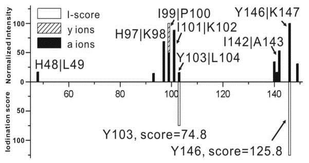 Figure 1. Fragment stack plot of RDD results for denatured Myoglobin, [DMyo•+15H]+15.
(Source: Sun, Q. Y. et al., 2010)
Protein iodination finds extensive application in various research areas, including molecular biology, immunology, and diagnostics. Here are some key applications:
Reference
For research use only, not for use in diagnostic procedures.
| Target | Cat. No. | Product Name | Size | Species Reactivity | Application | Detection Sample | |
| Tyrosine | DEIANS071 | Nitrotyrosine ELISA Kit | 96T | N/A | Quantitative | cell lysates, serum, plasma and purified proteins | Inquiry |
| DEIA3981 | Nitrotyrosine ELISA Kit | 96T | Human | Quantitative | Cell Lysate, Other biological fluids, Plasma, Serum, Urine | Inquiry | |
| DEIA087J | Nitrotyrosine ELISA Kit | 96T | Human | Quantitative | EDTA plasma, serum | Inquiry | |
| DEIA088J | Nitrotyrosine ELISA Kit | 96T | Human | Quantitative | stool, serum, EDTA plasma | Inquiry | |
| DEIA100J | Tyrosine ELISA Kit | 96T | Human | Quantitative | EDTA plasma, serum | Inquiry | |
| DEIA-PYR064 | Tyrosine Colorimetric Assay Kit | 100T | Quantitative | serum, plasma, urine, other body fluids | Inquiry | ||
| tyrosine hydroxylase | DEIA-XYA1809 | Tyrosine Hydroxylase (Phospho-Ser8) ELISA Kit | 2 x 96T | Qualitative | cultured cells | Inquiry | |
| DEIA-XYA1806 | Tyrosine Hydroxylase (Phospho-Ser19) ELISA Kit | 2 x 96T | Qualitative | cultured cells | Inquiry | ||
| DEIA-XYA1807 | Tyrosine Hydroxylase (Phospho-Ser31) ELISA Kit | 2 x 96T | Qualitative | cultured cells | Inquiry | ||
| DEIA-XYA1808 | Tyrosine Hydroxylase (Phospho-Ser40) ELISA Kit | 2 x 96T | Qualitative | cultured cells | Inquiry | ||
| Trypsin | DEIA-NS2310-14 | Trypsin ELISA kit | 96T | N/A | Quantitative | Biological samples | Inquiry |
Loading ......
