Phage display is a molecular biology technique in which a foreign protein or peptide gene is physically linked to a phage coat protein gene that physically displays the protein on the surface of the phage and that carries the foreign gene product in its own genome. Phage display can be used for the production of antibodies, peptides, proteins and enzymes. Phage display is now a technology for discovery and development of peptide and protein drugs, protein-protein interactions, protein-DNA interactions, protein structure and function, and targeted delivery of gene therapy. As phage display technology continues to advance, its advantages have attracted increasing attention from research teams, leading to expanded and improved applications. Building on phage surface display, related techniques have emerged, including baculovirus and other animal virus display systems, as well as yeast surface display.
Creative Diagnostics provides high-quality antibodies, antigens, and various reagents related to phage display technology to support your research and development needs.
The DNA sequence to be studied is placed into a specific site in the phage genome which is coding for a coat protein. When the phage infects the bacterial host, the phage genes are expressed. The peptide or antibody fragment inserted is then displayed on the surface of the phage as a fusion protein. If the inserted sequences are randomized, a phage display library with over 1010 variants can be created and stored for long periods as DNA clones, without having to individually construct, express and purify each peptide or antibody fragment. The important characteristic of phage display is the physical linkage of the displayed molecule (phenotype) to the DNA that encodes it (genotype). The ability to link genotype and phenotype enables the amino acid sequence of particular binders to be rapidly identified by sequencing the inserted DNA in the phage genome.
1. Library Design: Construct a library containing millions or more DNA clones encoding peptides or antibody fragments that can be expressed on the phage surface.
2. Library Cloning: Clone the library into the phage genome (using classic vectors or phagemid systems) and verify functional expression to ensure peptides are displayed on the phage particles.
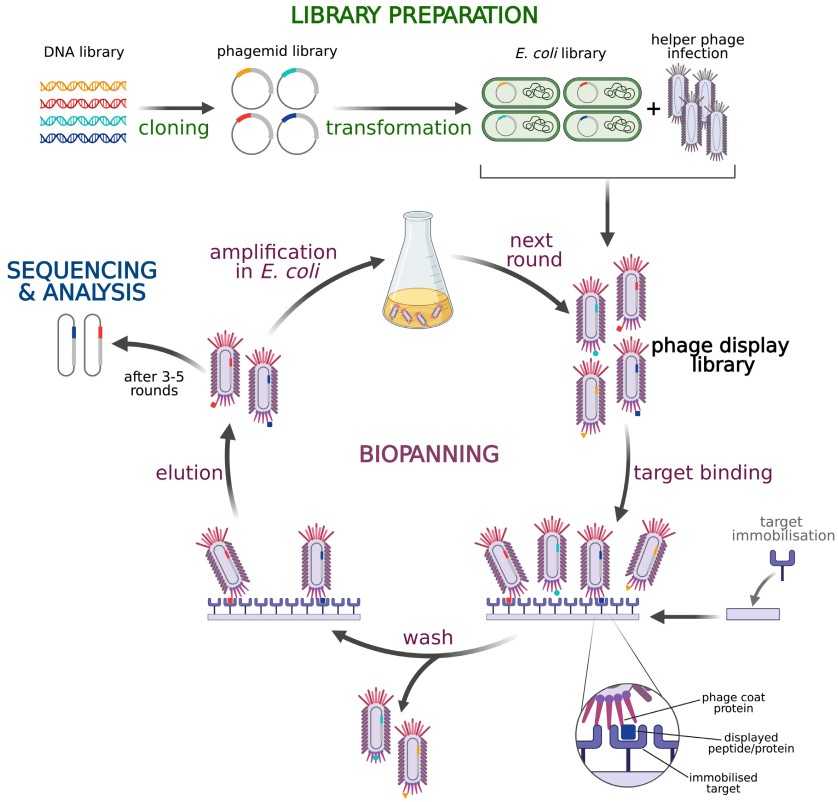 Figure 1. Library preparation and biopanning procedures based on phagemid and helper phage M13 pIII display
(Source: Jaroszewicz W, et al. 2022)
3. Selection Process (Biopanning): Use this step to easily identify specific phage particles.
4. Functional Validation and Mutant Library Construction: After confirming the function of peptides or proteins, develop diverse variant libraries through mutagenesis methods.
Can't find the proper solution?
Available ServicesPhage display libraries are large and diverse collections of phage clones, each with a random insertion of foreign DNA, resulting in each phage clone displaying a different molecule on the surface of the phage. The greatest benefit of using phage display libraries is that because of the high transformation efficiency of phages, a large number of phages can be screened in each round, making it more likely to find good binders. Other advantages are the low-cost amplification and ease of manipulation. However, there are some potential drawbacks, such as variable expression levels of certain peptide sequences in E. coli during screening, which may cause selection bias, and repeated library amplification can rapidly reduce phage diversity.
The diversity of phage display libraries is key to the success of the technology, and their construction strategies mainly fall into two categories:
Isolate B cells from immune or non-immune donors (for example, from human peripheral blood or spleen). After mRNA extraction and reverse-transcription to cDNA, antibody light and heavy chain gene fragments are PCR amplified and cloned into phage vectors to create single-chain variable fragment (scFv) or antigen-binding fragment (Fab) libraries.
 Protein-protein interactions
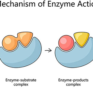
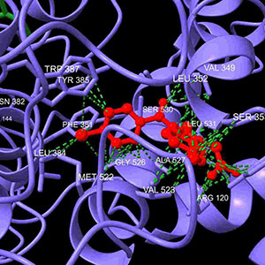
 Enzyme specificity and inhibitors
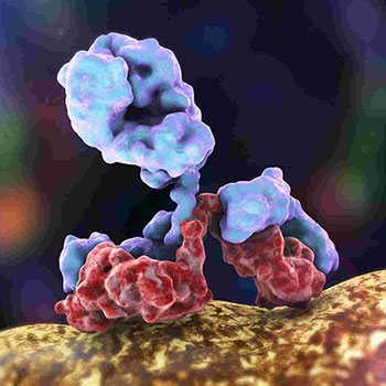
 Antibodies
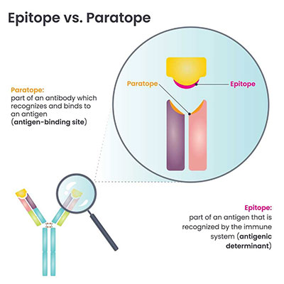
 Epitopes and mimotopes
 Receptors and G-proteins
Phage display of antibody fragments has been successfully used to generate antibodies targeting specific molecules, with broad applications in proteomics, targeted drug delivery, and analysis of intracellular processes. The main advantages of phage display for antibody generation are its speed and the avoidance of animal immunization, especially in humans.
Phage display is also used to select intrabodies—antibodies against intracellular targets. Intrabodies in the form of scFvs face challenges folding correctly in the reducing environment of the cytosol and nucleus, but highly diverse or optimized scFv phage libraries have successfully yielded functional intrabodies. These intrabodies are valuable for visualizing and modulating intracellular target functions.
Phage display is widely applied to discover antibodies against tumors and pathogens. One common approach is to use phage libraries against overexpressed molecules on tumor surfaces, or uniquely expressed on pathogen surfaces. Tumor-specific antibodies can be used to deliver disease-specific cell killing of imaging agents or cytotoxic conjugates (toxins, cytokines or radioisotopes). Targets unique to pathogens against which high-affinity antibodies can be selected can themselves be therapeutically neutralized by the antibodies.
Antibody phage display libraries can also be used to select anti-idiotypic antibodies that mimic antigen epitopes, which can be used in vaccine development. Filamentous phages themselves can be antigen carriers by fusing or crosslinking antigens to coat proteins to stimulate antibody responses.
One of the approaches of phage vaccines involves the foreign antigen display as a fusion protein on the surface of the phage capsid and the other approach involves direct conjugation of the antigen to the phage surface without any genome modification. Phages are very attractive as a carrier for vaccines as they can deliver the surface displayed antigens to the MHC-I or MHC-II molecules and lead to the cytotoxic T lymphocyte and antibody-mediated responses because the phages themselves are recognized as foreign by the host immune system and can be readily taken up by the antigen presenting cells. Also, since phages only infect the prokaryotes, they are considered to be safe for human use. The various phage families differ from one another in terms of their size, shape, and the surface proteins employed for antigen display.
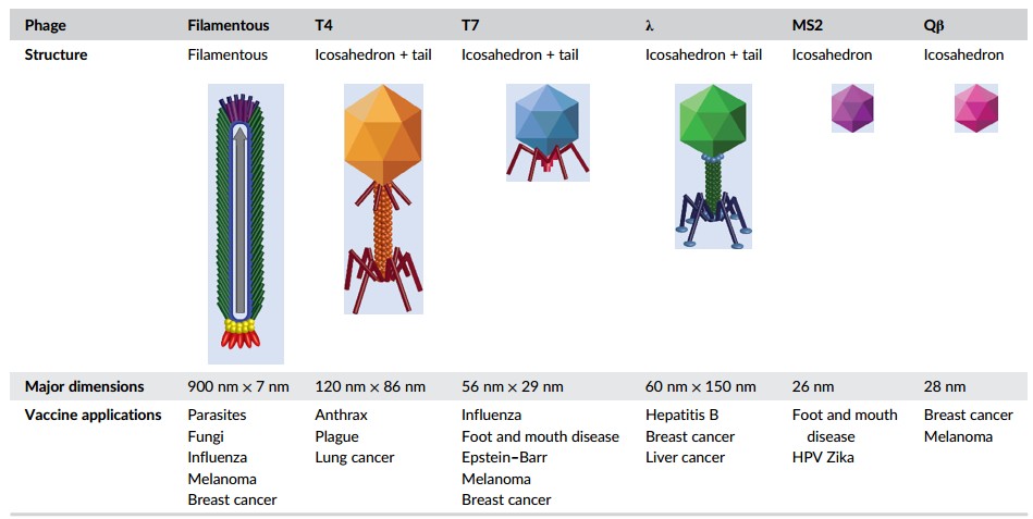 Figure 2. Structure and application of families of phages used as vaccine platforms. (Source: Hess KL, et al. 2019)
References