
Inquiry Basket
 Loading ......
Malaria is a mosquito-borne parasitic disease causing high morbidity and mortality primarily in infants and young children. People with malaria often experience fever, chills, and flu-like illness. Left untreated, they may develop severe complications and die. In 2020 an estimated 241 million cases of malaria occurred worldwide and 627,000 people died. Malaria is caused by Plasmodium, which was first described in the year 1885 and now contains about 200 species. In humans, malaria is caused by six Plasmodium species: P. falciparum, P. malariae, P. ovale curtisi, P. ovale wallikeri, P. vivax and P. knowlesi. Among these, P. falciparum and P. vivax are the major causative agents of malaria.
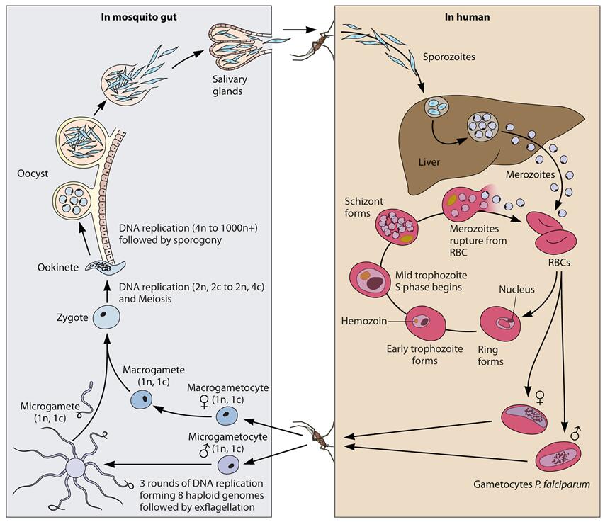 Fig. 1 The Plasmodium life cycle (Lee AH, et al. 2014)
Plasmodium parasite always has two hosts in its life cycle: a dipteran insect host (for example, mosquitos) and a vertebrate host. The life cycle of Plasmodium species involves some different stages in both the two kinds of hosts, including sporozoites, merozoites, gametocytes and hypnozoites in some species. Plasmodium parasites exist in the salivary gland of infected mosquitoes as sporozoite form. Sporozoites are injected into the vertebrate host along with the saliva when the infected mosquito bites the vertebrate host. The sporozoites join in the blood stream and then are transported to the liver, where they invade and replicate within liver cells (or hepatocytes). Newly produced parasite (merozoite form) return to the blood and start to invade red blood cells. Merozoites grow first to a ring-shaped form and then turn to a larger trophozoite form. Trophozoites then mature to schizonts which divide several times to produce new merozoites. The infected red blood cells eventually burst, allowing the newly produced merozoites return back to the blood and invade new red blood cells. Most merozoites continue this replicative cycle while some of them differentiate into male or female sexual forms called gametocytes. These gametocytes exist in the blood circling system until a mosquito feeds on the infected vertebrate host, taking up them along with the blood. Gametocytes develop into male and female gametes in the salivary gland of infected mosquitoes and form a zygote. Zygotes will finally develop into sporozoites and continue the life cycle.
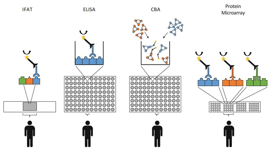 Fig. 2 The Malaria diagnostic platform, antibody detection (Hoogen LVD & Drakeley C. 2015)
Malaria is conventionally diagnosed by microscopic examination of stained blood films, other tests including serology, PCR, and drug-resistance testing. Diagnosis of malaria using serological methods is usually based on the detection of antibodies against malaria parasites, using either indirect immunofluorescence (IFA) or enzyme-linked immunosorbent assay (ELISA). Serology does not detect current infection but rather measures past exposure. IFA is a reliable serologic test for malaria because of its highly sensitive and specific. Following infection with any Plasmodium species, specific antibodies are produced within 2 weeks of initial infection, and persist for 3-6 months after parasite clearance. IFA uses specific antigen or crude antigen prepared on a slide, and quantifies both IgG and IgM antibodies in patient serum samples. Titers > 1 : 20 are usually deemed positive, and titers > 1 : 200 can be classified as recent infections.
Loading ......
