

 Bioprocess Impurity Analysis Protein A
Inquiry Basket
 Loading ......
Protein A, Protein G, and Protein L are proteins isolated from the surface of bacterial species. The affinity chromatography of them is a commonly used method for the purification of therapeutic antibodies. Therapeutic antibodies have a wide range of applications ranging from drugs against cancer and autoimmune diseases to labeling and detection of virulence factors. In the biomanufacturing process of therapeutic antibodies, in addition to high product yields, strict critical quality attributes (CQAs) have to be reached, among which purity is an important indicator. Protein A, Protein G, and Protein L have been found as impurities that may leach during the affinity chromatography processes of therapeutic antibody manufacturing. Apart from that, due to the different properties of Protein A, Protein G, and Protein L, they are also currently used in other applications such as vaccine development and purification of special therapeutic antibodies.
Protein A is often coupled to other molecules such as a fluorescent dye, enzymes, colloidal gold or radioactive iodine without affecting the antibody binding site. Examples including protein A–gold (PAG) stain is used in immunogold labelling, fluorophore coupled protein A for immunofluorescence, and DNA docking strand coupled protein A for DNA-PAINT imaging. It is also widely utilized coupled to magnetic, latex and agarose beads. Besides, protein A is also a key factor in the generation of an effective S. aureus vaccine. The SpA encompasses five Immunoglobulin binding domains (IgBDs) that associate with the Fcγ domain to block phagocytosis, causing the inability to generate an efficacious humoral immune response. Due to this reason the development of an anti-S.aureus vaccine has been inhibited. Thus, the disruption of SpA's role as a virulence factor may be a key step in the generation of an effective S. aureus vaccine.
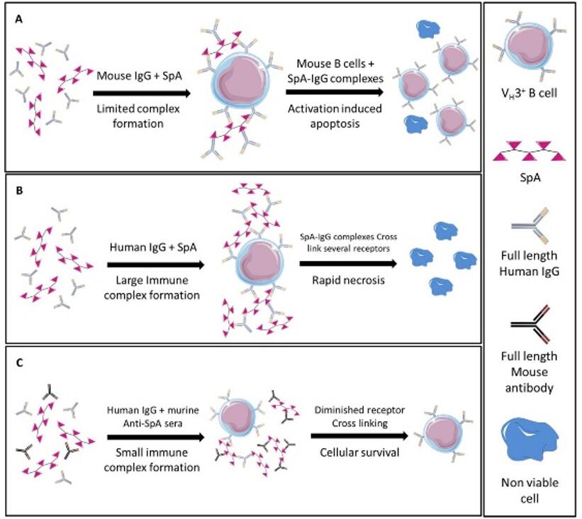 Fig 1. Proposed mechanism of action for SpA-induced toxicity
Protein G is first isolated from a human group G streptococcal bacterium. The gene structure and protein-binding properties of Streptococcal protein G (SPG) are similar to protein A, but SPG is with a greater affinity to most mammalian IgGs, particularly for certain subclasses, including human IgG3, mouse IgG1, and rat IgG2a. However, unlike protein A, SPG does not bind to human IgM, IgD, or IgA. So SPG may be used for the purification of mammalian monoclonal and polyclonal IgGs, which do not bind well to protein A, and has an important role in the conjugation of antibodies with sensitive immunolabeled molecules. Besides, recombinant Protein G, either labeled with a fluorophore or a single-stranded DNA strand, was used as a replacement for secondary antibodies in immunofluorescence and super-resolution imaging. Apart from that, the use of purified rSPG conjugates may be vital for the diagnosis of certain diseases, such as autoimmune diseases, since IgG3 is overexpressed in numerous autoimmune diseases (represents ≥45% of autoimmune antibodies).
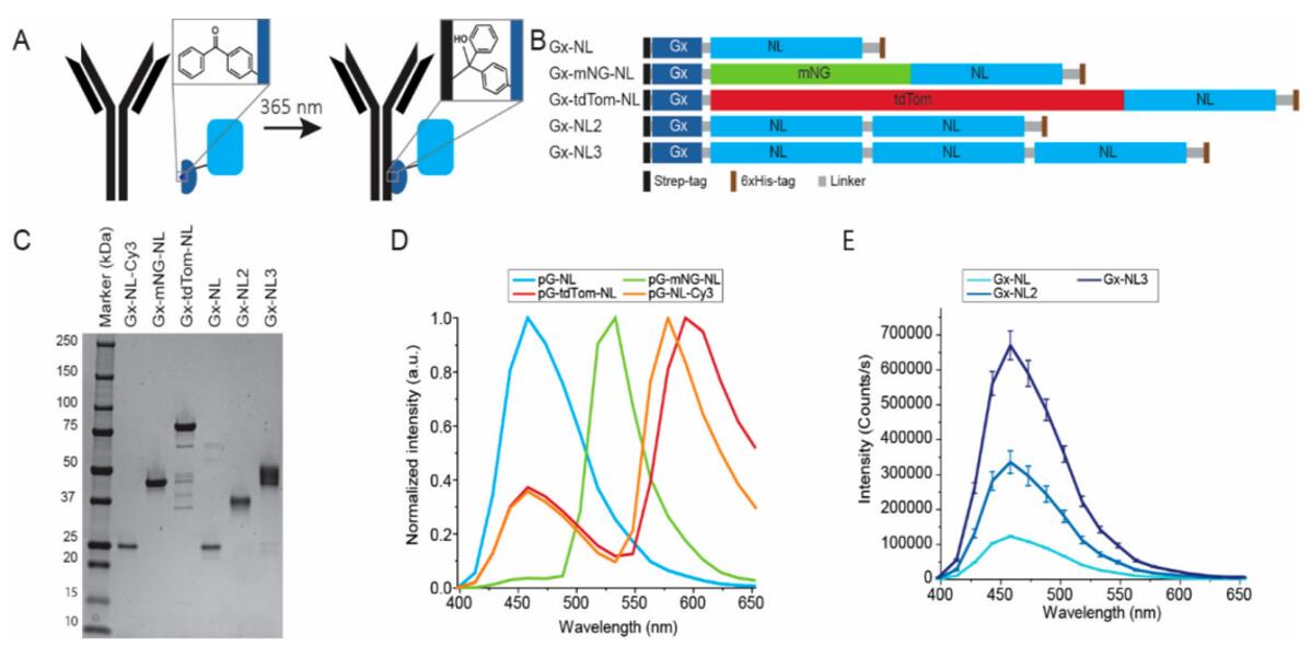 Fig. 2 Development of bioluminescent antibodies using Gx-NL fusion proteins (NanoLuc luciferase genetically fused to a protein G).
Protein L is an immunoglobulin (Ig)-binding protein that binds to the variable light chains (kappa chain) of Ig without interfering with antigen binding site, because of which it can bind to single-chain antibody fragments (scFv) and Fab fragments. Protein L method has already been established for the purification of fab fragments. Its ability of bind to scFv also makes it a convenient reagent to detect the cell surface expression of CARs in adoptive immunotherapy research. A further application of protein L in the DSP is the separation of homodimers from heterodimeric bispecific antibodies (bsAbs). Homodimers are commonly found byproducts during the recombinant production of asymmetric IgG-like bsAbs. Their monitoring and removal generally pose great challenges to analytical and purification process, respectively, as they share high similarity in physicochemical properties with the target heterodimeric bsAb. It has been reported that after Protein L affinity chromatography using an additive of Arg·HCl in the elution buffer at pH3.0, the aggregate amount can be reduced from 66.5% in the cell culture supernatant to 7.1%.
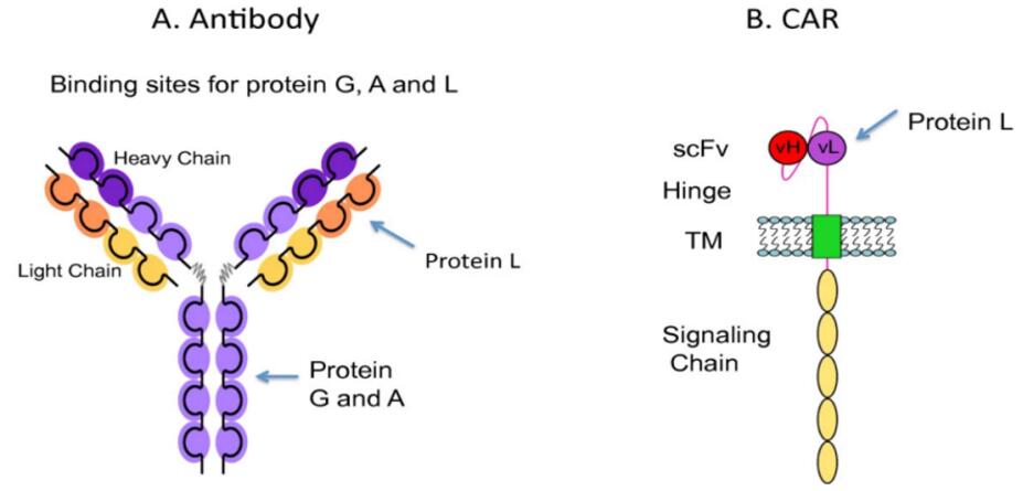 Fig 3. The binding sites of Protein A, Protein G, and Protein L to the heavy and light chain regions of the antibody
Creative Diagnostics offers highly specific antibodies that detect Protein A, Protein L, and protein G in several scientific applications, including Western Blot, ELISA, Immunohistochemistry, Immunoprecipitation, and Immunocytochemistry. Among these includes a set of Chicken antibodies, which have biochemical advantages over mammalian antibodies, resulting in increased sensitivity and a decreased background in immunological assays. They have also been found with higher titer and better stability. All our antibodies are fully verified. If you cannot find the antibody you're looking for, contact us to develop customized antibodies for specific applications.
| Cat No. | Product Name | Applications | Inquiry |
| DPATB-H82918U | Magic™ Chicken Anti-Protein G polyclonal antibody | WB, ELISA | Inquiry |
| DPATB-H82918 | Magic™ Chicken Anti-Protein G polyclonal antibody [HRP] | WB, ELISA | Inquiry |
| DPAB-DC4773 | Rabbit Anti-Protein G polyclonal antibody | ELISA | Inquiry |
| DPATB-H83138 | Rabbit Anti-Protein G polyclonal antibody [Biotin] | WB, ELISA | Inquiry |
| DPATB-H83139 | Rabbit Anti-Protein G polyclonal antibody [Texas Red] | FC, IA | Inquiry |
| DPABH-29366 | Rabbit Anti-Protein G polyclonal antibody | IHC-P, WB | Inquiry |
| DAG209 | Recombinant Protein G | ELISA | Inquiry |
| DAG-T1430 | Recombinant Protein G [Gold] | IA | Inquiry |
| DAG677 | Recombinant Strepococcus Protein G | IP, ELISA | Inquiry |
| DAG3602 | Recombinant Streptococcus Protein G | IA | Inquiry |
| DAG678 | Recombinant Strepococcus Protein G [HRP] | ELISA | Inquiry |
| DEIASL150 | Protein G ELISA Kit | Inquiry |
| Cat No. | Product Name | Applications | Inquiry |
| DPATB-H83614 | Magic™ Chicken Anti- Protein L Polyclonal antibody | WB, ELISA | Inquiry |
| DPATB-H83619 | Magic™ Chicken Anti- Protein L Polyclonal antibody [HRP] | WB, ELISA | Inquiry |
| DPATB-H83617 | Magic™ Chicken Anti- Protein L Polyclonal antibody [FITC] | ELISA, LF, WB | Inquiry |
| DPATB-H8361B | Magic™ Chicken Anti- Protein L Polyclonal antibody [Biotin] | WB, ELISA | Inquiry |
| DPAB-DC4664 | Rabbit Anti-Protein L Polyclonal antibody | WB | Inquiry |
| DAG679 | Recombinant Protein L | IP | Inquiry |
| DAG2476 | Recombinant Protein L | IA | Inquiry |
| DAG4610 | Protein L [HRP] | WB, ELISA, DB | Inquiry |
| DEIASL153 | Protein L Ligand Leakage ELISA | Inquiry |
Loading ......
