
Inquiry Basket
 Loading ......
Neuroendocrine tumors are tumors that originate from neuroendocrine cells. Neuroendocrine cells are a large group of cells in the body with neuroendocrine phenotypes that can produce various hormones. Neuroendocrine cells are distributed throughout the body, so neuroendocrine tumors can occur in any part of the body, but the most common are neuroendocrine tumors in the digestive system such as the stomach, intestines, and pancreas, accounting for about 2/3 of all neuroendocrine tumors. The incidence rate of neuroendocrine tumors in European and American populations is also very high. The incidence rate has increased more in the past 30 years. Compared with other tumors, the incidence rate of neuroendocrine tumors has increased more rapidly.
 Figure 1. Neuroendocrine Tumors.
When neuroendocrine cells undergo genetic mutations leading to uncontrolled proliferation, NETs will develop. They can occur anywhere these cells are present, but the most common primary site is:
Most neuroendocrine tumors are sporadic, and their exact etiology is currently unclear. However, a small portion of neuroendocrine tumors are related to genetic factors, involving the loss and mutation of certain genes, such as multiple endocrine adenomas (MEN) and von Hippel Lindau syndrome (VHL syndrome).
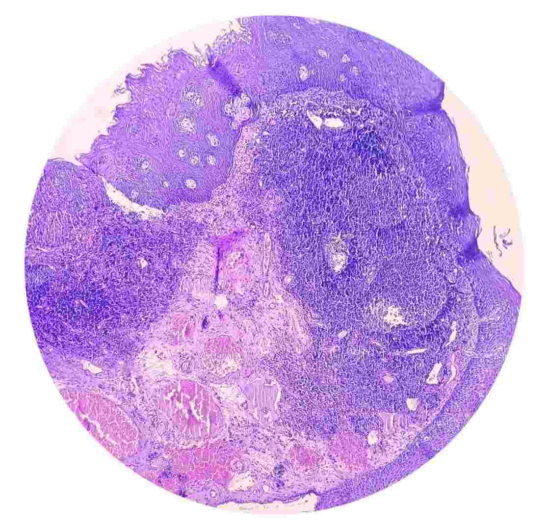 Figure 2. Etiology.
Neuroendocrine tumors are classified into two categories: non functional (about 80%) and functional (about 20%) based on whether the tumor has hormone secretion function and whether there are clinical symptoms caused by hormones. Non functional gastrointestinal pancreatic neuroendocrine tumors are mainly characterized by non-specific gastrointestinal symptoms or local tumor occupying symptoms, such as progressive swallowing difficulties, abdominal pain, bloating, diarrhea, abdominal masses, jaundice, or black stools; Functional gastrointestinal and pancreatic neuroendocrine tumors are mainly characterized by clinical symptoms caused by the secretion of biologically active hormones, such as skin flushing, sweating, asthma, diarrhea, hypoglycemia, refractory gastrointestinal ulcer, diabetes, etc. Functional gastrointestinal and pancreatic neuroendocrine tumors are mainly pancreatic neuroendocrine tumors, including insulinoma, somatostatin tumor, glucagon tumor, gastrinoma, etc.
| Disease Examination | Details |
| Tumor marker examination | There is a very important tumor marker for neuroendocrine tumors, called chromogranin A (CgA), which is currently the most valuable universal marker for neuroendocrine tumors (whether functional or non functional). By detecting serum or plasma levels of chromaffin A, it is possible to indicate whether patients have neuroendocrine tumors, track their treatment response, and even evaluate their prognosis. The diagnostic sensitivity and specificity of serum or plasma chromaffin A detection are between 60% and 95%. In addition to the universal tumor marker chromochrome A, functional neuroendocrine tumors can also be diagnosed by detecting the specific hormones they secrete. For example, gastrinoma can detect serum gastrin levels, and insulinoma can detect serum insulin levels. |
| Imaging Examination | Various imaging examinations, including endoscopy, endoscopic ultrasonography, ultrasound CT, PET-CT, MRI, Somatostatin receptor imaging (SRS) and other techniques are important tools for localizing and diagnosing neuroendocrine tumors. |
| Pathological Examination | The final diagnosis of neuroendocrine tumors relies on pathological examination. The key points of pathological diagnosis of neuroendocrine tumors include: firstly, determining whether the tumor is a neuroendocrine tumor through immunostaining of neuroendocrine markers such as synaptophysin (Syn) and chromochrome A, and secondly, determining the grading of the tumor based on its proliferative activity. The proliferative activity of tumors is evaluated by the number of mitotic figures or Ki-67 positivity index. According to the proliferative activity of tumors, gastrointestinal and pancreatic neuroendocrine tumors are classified into G1 (low grade, with 1/10 of the number of nuclear fission figures in the high-power field or Ki-67 index ≤ 2%), G2 (medium grade, with 2-20 of the number of nuclear fission figures in the high-power field or Ki-67 index of 3% -20%), and G3 (high grade, with more than 20/10 of the number of nuclear fission figures in the high-power field or Ki-67 index>20%). |
Diagnosis of neuroendocrine tumors is based on corresponding clinical manifestations, tumor marker detection, imaging examination, and pathological examination. The complete diagnostic content includes tumor location, grading, staging, and functional status.
The treatment methods for neuroendocrine tumors include endoscopic surgery and surgical treatment, radiation intervention therapy, radionuclide therapy, chemotherapy, biological therapy, molecular targeted therapy, etc. The choice of treatment method depends on the grading, staging, location of occurrence, and whether the tumor has hormone secretion function. For localized tumors, radical surgery can be performed to remove them; For patients with advanced tumors, some can also receive palliative treatment through surgical tumor reduction surgery; For patients with only liver metastases, local treatment for liver metastases can be chosen, including various ablations, hepatic artery embolization, radioactive particle implantation, and even liver transplantation; For metastatic neuroendocrine tumors, radiolabeled somatostatin analogs can also be used for peptide receptor-mediated radionuclide therapy, abbreviated as PRRT therapy (PRRT).
Neuroendocrine tumors were once considered obscure and untreatable, but now they are at the forefront of oncology innovation. In the past two decades, significant progress has been made in diagnostic accuracy, the development of effective biological therapies (SSA), and the emergence of targeted radiation therapy (PRRT). These developments have transformed NETs into chronic, manageable diseases for many patients, significantly improving their quality of life and overall survival rates. However, challenges still exist, particularly in early detection and management of treatment resistance. The ongoing research continues to uncover the complex genetics of NETs, paving the way for more personalized and effective treatments.
| Cat. No. | Product Name | Host | Isotype | Application | |
| CPBT-66665SH | Anti-CHGA (aa 1-15) polyclonal antibody | Sheep | IgG | ELISA, IHC-P | Inquiry |
| DPABB-JX57P | Chicken anti-Rat Peripherin polyclonal antibody | Chicken | IgG | ELISA, WB, IF | Inquiry |
| CABT-Z197R | Recombinant Rabbit Anti-Human CDX2 Monoclonal Antibody, clone CQ7134 | Rabbit | IgG | IHC-P | Inquiry |
| DPAB-TJ020 | Anti-Pancreatic cell line mucin polyclonal antibody | Rabbit | IgG | IHC | Inquiry |
| DMABB-JX143 | Mouse Anti-Human Chromogranin A Monoclonal Antibody, clone 3H9 | Mouse | IgG2b | ELISA, WB, IP | Inquiry |
| DMABB-JX144 | Mouse Anti-Human Chromogranin B Monoclonal Antibody, clone 2F22 | Mouse | IgG1 | ELISA, WB | Inquiry |
| DMAB5335MH | Anti-CHGA monoclonal antibody, clone ML3I11 | Mouse | IgG1 | IHC | Inquiry |
| DCABH-4960 | Rabbit Anti-NOTCH4 monoclonal antibody, clone KN212-16 | Rabbit | IgG | WB, FC | Inquiry |
| DCABH-9081 | Rabbit Anti-NOTCH1 monoclonal antibody, clone TK316 | Rabbit | IgG | WB, ICC/IF, IHC, IP, FC | Inquiry |
| DCABH-8870 | Anti-NCAM1 monoclonal antibody, clone 844/NFN-299 [FITC + PE] | Mouse | FC | Inquiry |
| Cat. No. | Product Name | Size | Species Reactivity | Application | Detection Method | |
| DEIA4545 | Mouse/Rat Corticotropin Releasing Factor ELISA Kit, Sensitive, Extraction-free | 96T | Mouse, Rat | Quantitative | sELISA | Inquiry |
| DEIA-JY2408 | Serotonin ELISA kit | 96T | Human | Quantitative | ELISA | Inquiry |
| Cat. No. | Product Name | Size | Target | Species | |
| DAG-WT1248 | Recombinant Human NSE [His] | 100 g | NSE | Human | Inquiry |
| DAG-T1014 | Human NSE | 50 g | Human | Inquiry | |
| DAG-WT1130 | Inactivated Seneca Valley virus (SVV) | 1 mL | SVA | Inquiry | |
| DAG-WT2502 | Neuron-specific enolase (NSE) control | 0.5 mL, 1.0 mL | NSE | Inquiry |
Loading ......
