
Inquiry Basket
 Loading ......
Breast cancer is a heterogeneous disease with distinct pathological and molecular properties, as well as clinical manifestations. Breast cancer can be categorized into five main intrinsic molecular subtypes based on molecular features: Luminal A, Luminal B, HER2-enriched, Basal-like, and Claudin-low. Invasive ductal carcinoma is the most prevalent malignant breast tumor, accounting for 75% of cancer cases and having a high lymphatic metastatic risk. Initial breast cancers can spread through lymphatic or blood vessels, resulting in distant metastasis, particularly to bones, which can cause discomfort, mobility difficulties, neurogenic disorders, hypercalcemia, and pathological fractures. The later the diagnosis, the more likely metastasis, which can occur in the brain, bone marrow, liver, lymph nodes, and lung.
Among women, breast cancer is the most common malignancy, accounting for approximately 25% of newly diagnosed cancer cases worldwide. In the United States, about 13% of women develop breast cancer. The incidence of breast cancer varies globally, with the highest rates in Australia, New Zealand, most parts of Europe, and North America, intermediate rates in South America and Eastern Europe, and the lowest rates in most parts of Asia and Africa. The etiology of breast cancer is complex and multifaceted, involving genetic, environmental, and lifestyle factors. The incidence is associated with multiple factors, including gender, age, estrogen levels, family history, gene mutations (such as BRCA1 and BRCA2 mutations), and unhealthy lifestyle choices, among others. Despite the increasing incidence of breast cancer globally, mortality rates have declined due to improved screening and treatment methods over recent decades.
Breast cancer treatment is multidisciplinary, including local treatments (surgery and radiotherapy) and systemic therapies. Systemic treatments include endocrine therapy for hormone receptor-positive disease, chemotherapy, anti-HER2 therapy for HER2-positive disease, bone-stabilizing agents, poly (ADP-ribose) polymerase inhibitors for BRCA mutation carriers, and recent immunotherapies. Despite significant advances in breast cancer treatment, local and distant recurrences remain major threats. About 20-30% of early-stage breast cancer patients eventually develop distant metastatic disease recurrence. Moreover, achieving equitable access to treatment advancements globally remains a future challenge in breast cancer care.
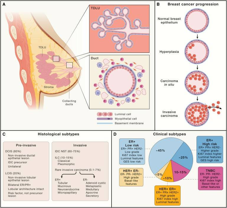 Figure 1. Anatomy of the Breast and Pathological Classification of Breast Cancer Tissues
(Source: Nolan E, et al. 2023)
Tumor markers (TMs) are a group of molecules with different biochemical structures and functions (such as hormones, enzymes, cancer proteins, etc.), directly or indirectly synthesized and released into the bloodstream by the presence of tumors. Tumor markers are useful in various aspects of cancer patient management, particularly in diagnosis and monitoring treatment response. An ideal tumor marker should have high specificity and sensitivity, being secreted into the blood at measurable concentrations following malignant transformation, with results reflecting the tumor's location. However, to date, no tumor marker exists with 100% specificity and 100% sensitivity.
In breast cancer, the most widely used serum tumor markers are Cancer Antigen 15-3 (CA 15-3) and Carcinoembryonic Antigen (CEA). These markers are frequently released during metastatic disease and their release precedes clinical symptoms and detection by medical imaging. Studies have shown that preoperative serum levels of CA 15-3 and CEA are significantly associated with tumor size, axillary lymph node metastasis, and advanced staging.
CA 15-3 is a protein with a high molecular weight (300-450 KDa) that belongs to the transmembrane receptor MUC-1 and possesses cell adhesion properties. MUC-1 can be found on the apical plasma membrane of normal secretory epithelial cells. However, following malignant transformation, MUC-1 may be overexpressed on both the membrane surface and the cytoplasm. CA 15-3 identifies a soluble version of MUC-1 protein and is the most often utilized blood marker in breast cancer patients. In breast cancer patients, CA 15-3 expression in tumor cells triggers the production of antibodies (anti-CA 15-3) as part of the immune response. Approximately 80% of patients with metastatic breast cancer have CA 15-3 leakage from cancer cell surfaces into the blood. Thus, this glycoprotein antigen is the most widely used serum tumor marker in breast cancer. The reference range for serum CA 15-3 is less than 30 U/mL.
 Figure 2. CA 15-3 Levels Comparison in Healthy Individuals and Breast Cancer Patients
(Source: Siddiqua J, et al. 2010)
CA 15-3 is essential for detecting and monitoring breast cancer. It contributes to determining the amount of breast cancer dissemination, particularly bone metastases. Elevated CA 15-3 levels indicate metastases, particularly bone metastasis. CA 15-3 can be considered a useful predictor of bone metastase prognosis in relevant patients.
To detect CA 15-3, several analytical techniques can be utilized, including enzyme-linked immunosorbent assay (ELISA), fluorescence immunoassay, electrochemiluminescence, and radioimmunoassay. However, these techniques have certain disadvantages, including expensive costs, the requirement for experienced personnel, lengthy analysis periods, and sophisticated labor-intensive testing procedures.
CEA, a high-molecular-weight glycoprotein from the immunoglobulin family, was originally isolated from colon cancer and embryonic colonic mucosa in 1965. CEA is expressed in a variety of tissues, including the stomach, colon, pancreas, and lungs. The CEA family is separated into three branches: CEA-related cell adhesion molecules (CEACAM), pregnancy-specific glycoproteins (PSG), and pseudogenes. CEA plays a critical role as a regulator of tumor cell proliferation and differentiation, and its overexpression leads to the progression of many epithelial cancers and immune dysfunctions. Although CEA is more widely used in the management of colorectal cancer, it also holds significant importance in the management of breast cancer.
CEA levels are significantly higher in breast cancer patients than in healthy individuals. Although CEA's sensitivity is lower than that of CA 15-3, it remains a valuable tumor marker in certain circumstances. Compared to CA 15-3, CEA is more suitable for preoperative and postoperative monitoring. Techniques for detecting CEA include enzyme-linked immunosorbent assay (ELISA), electrochemiluminescence, radioimmunoassay, and immune biosensor.
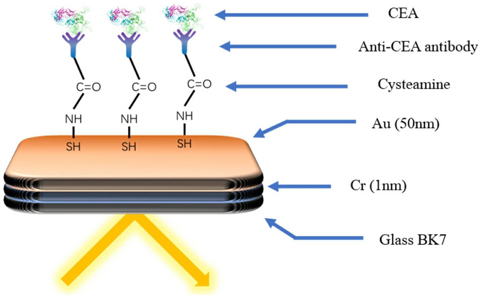 Figure 3. Diagram of an Immunobiosensor for CEA Detection
(Source: Niu J Hong R, et al. 2022)
In summary, breast cancer is the most common malignancy among women worldwide, and despite continuous improvements in treatment methods, many challenges remain. CA 15-3 and CEA are the most widely used serum tumor markers in breast cancer, playing crucial roles in early and metastatic diagnosis, treatment monitoring, and prognosis evaluation. Further research and improvements in detection technologies can enhance the sensitivity and specificity of these markers, better serving the management and treatment of breast cancer patients. If your laboratory is conducting or planning to conduct breast cancer detection and research or in need of tumor markers, Creative Diagnostics offers a range of high-quality cancer antigens (including CA15-3) and ELISA kits. All our products undergo extensive quality control validation to ensure the reliability of your experiments. Please visit our product page to place an order, and they will arrive quickly and safely at your laboratory bench.
References
| Target | Cat. No. | Product Name | Size | Application | Detection Sample | Column8 | |
| CA 15-3 | DEIA2145 | CA15-3 ELISA Kit | 96T | Human | Quantitative | Serum or plasma | Inquiry |
| Target | Cat. No. | Product Name | Host | Isotype | Application | |
| CA15-3 | DCABH-20038 | Mouse Anti--Human CA15-3 monoclonal antibody, clone Y20 | Mouse | IgG | IHC, IB, IA, ELISA(Cap) | Inquiry |
| CA15-3 | DCABH-20039 | Mouse Anti--Human CA15-3 monoclonal antibody, clone Y219 | Mouse | IgG | IHC, IB, IA, ELISA(Det) | Inquiry |
| CA15-3 | DMAB27451 | Anti-CA15-3 monoclonal antibody, clone EG4 | Mouse | IgG1 | IHC | Inquiry |
| CA15-3 | DMAB5364MH | Anti-CA15-3 monoclonal antibody, clone ECNN2 | Mouse | IgG1 | IHC | Inquiry |
| Target | Cat. No. | Product Name | Expression System | Tag/Conjugate | Application | |
| CA15-3 | DAG700 | Human Breast Cancer Antigen (CA 15-3) | Human fluids | Unconjugated | ELISA, Controls | Inquiry |
| CA15-3 | DAGF-212 | Human milk derived MUC1 (CA15-3) antigen | Human fluids | Unconjugated | Inquiry |
| Target | Cat. No. | Product Name | Host | Isotype | Application | |
| CEA | CABT-L2986 | Mouse Anti-Human CEA monoclonal antibody, clone JID654 | Mouse | IgG | IHC | Inquiry |
| CEA | CABT-L2017 | Rabbit Anti-Human CEA Polyclonal Antibody | Rabbit | IgG | WB | Inquiry |
| CEA | CABT-Z665H | Human Anti-Human CEA (Arcitumomab) Monoclonal Antibody, clone Arcitumomab [Biosimilar] | Human | IgG1, κ | IHC-P, FC | Inquiry |
| Target | Cat. No. | Product Name | Expression System | Tag/Conjugate | Application | |
| CEA | DAGA-773 | CEA protein (>95%) | Human liver carcinoma cells | Unconjugated | N/A | Inquiry |
| CEA | DAG-WT2581 | Carcinoembryonic antigen (CEA) control | N/A | Unconjugated | Immunoassays | Inquiry |
| Target | Cat. No. | Product Name | Size | Application | Detection Sample | |
| CEA | DEIA-NS2307-76 | Human CEA(Carcinoembryonic Antigen) ELISA Kit | 96T | Human | Quantitative | Inquiry |
Loading ......
