Green fluorescent protein (GFP) is the first fluorescent protein discovered. It is a β-barrel protein derived from the crystal jellyfish Aequorea Victoria. GFP has a molecular weight of about 27 kDa and is a single-chain polypeptide consisting of 238 amino acids. It has a fluorescence emission wavelength in the green part of the visible spectrum due to a chromophore formed by the maturation reaction of three specific amino acids (Ser65, Tyr66, and Gly67) in the center of the protein. When initially discovered, one of the most surprising aspects of GFP was that the chromophore formed spontaneously, without additional cofactors, substrates, or enzymatic activity, and it only required the presence of oxygen during maturation. This means that the protein can be extracted directly from A. victoria and expressed in any organism while still remaining fluorescent.
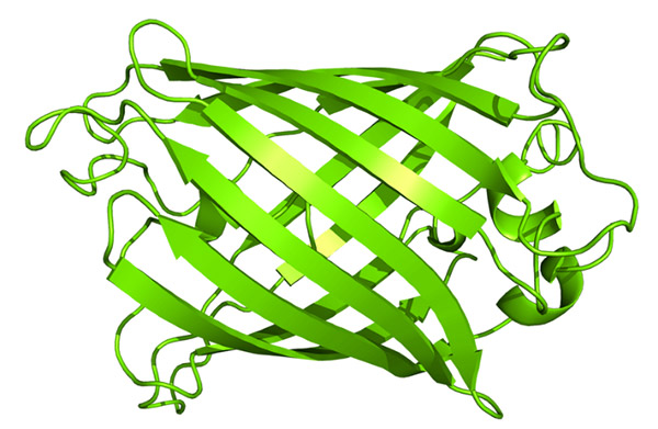 Fig. 1 Cartoon rendering of a 1.9 Å crystal structure of the GFP of A. victoria. (Schmid B, 2020)
The main advantage of GFP over traditional fluorescent dyes is that it is non-toxic and can be expressed in living cells, enabling the study of dynamic physiological processes. In addition, the high-resolution crystal structure of GFP enables scientists to manipulate its protein structure to increase its color and range of applications.
The discovery and development of GFP have led to significant advances in biological research. GFP's unique properties have made it a valuable tool for visualizing and tracking cellular and molecular processes in real time. GFP is often used as a reporter protein to track the expression and localization of proteins of interest in living cells. Its ability to emit light without a substrate or cofactor has made it particularly useful for studying dynamic cellular processes, such as protein trafficking, signal transduction, and gene expression.
One of the most significant applications of GFP in biological research is its use as an imaging and tracking tool for cellular processes. GFP can be fused to proteins of interest, allowing researchers to visualize their expression and localization in living cells. For example, GFP can be fused to a protein that localizes to the cell membrane, allowing researchers to track its movement and distribution in real time. Similarly, GFP can be fused to transcription factors, enabling researchers to track their localization and activity in response to different stimuli.
GFP has also been used in protein-protein interaction studies. Interactions that undergo conformational changes between two proteins or between two domains of a protein may be studied by fusing GFP individually to each protein or domain of interest. This technique, known as fluorescence resonance energy transfer (FRET), has been used to study protein-protein interactions in a variety of biological systems, including neurons and cancer cells.
Expression vectors such as plasmids often contain GFP as a marker to help determine which cells have successfully taken up the plasmid. This can be an alternative to antibiotic options. This type of plasmid may produce GFP under the control of another promoter of the gene of interest, or express GFP from the same transcript as the gene of interest, but after the internal ribosome entry site (IRES).
Fluorescence-activated cell sorting (FACS) is a type of flow cytometry that separates a mixture of cells into distinct populations based on a fluorescent signal. FACS can be used to separate GFP-expressing cells from non-GFP-expressing cells.
Another important application of GFP in biological research is gene expression analysis. GFP can be used as a reporter gene to monitor the expression of specific genes in living cells. For example, GFP can be fused to a promoter sequence of a gene of interest, allowing researchers to monitor its expression in real-time. This technique, known as GFP reporter assay, has been used to study gene expression in a variety of biological systems, including bacteria, yeast, and mammalian cells.
GFP-based fluorescent biosensors have been designed to detect a variety of intracellular conditions, including ion (such as Ca2+) concentration and pH, using a series of strategies, such as FRET, calmodulin, etc.
In addition, GFP is also used to identify specific cell populations in drug screening, visualize micrometastasis in nude mice in cancer research, serve as the reporter of DNA double-strand break repair, and mark pathogenic intracellular microorganisms to visualize host/pathogen interaction.
As a leading biotechnology company, Creative Diagnostics offers a range of high-quality GFP products, including antibodies, proteins, and ELISA kits, that are validated for use in a variety of key applications, such as immunohistochemistry, western blot, flow cytometry, immunocytochemistry, ELISA, and immunoprecipitation.
References