
Inquiry Basket
 Loading ......
Bacillus anthracis is capable of causing anthrax, which is a serious threat to human life and health. The bacterium is a non-motile, gram-positive bacterium with an envelope that contains two forms, including the vegetative form, which is active, and the spore form, which is biologically inert. Bacteria in the spore form are resistant to heat, cold, radiation, and disinfectants and can therefore survive for long periods of time in a variety of environments. It is endemic in sub-Saharan Africa, the Middle East, Central and South-West Asia, and Central and South America. There are also sporadic cases in North America and Europe, with the majority occurring in Eastern and Southern Europe. Natural human infections with Bacillus anthracis usually follow animal outbreaks and are associated with the slaughter and consumption of dead anthrax-diseased animals in countries with food shortages, inadequate veterinary inspections, or low vaccination coverage.
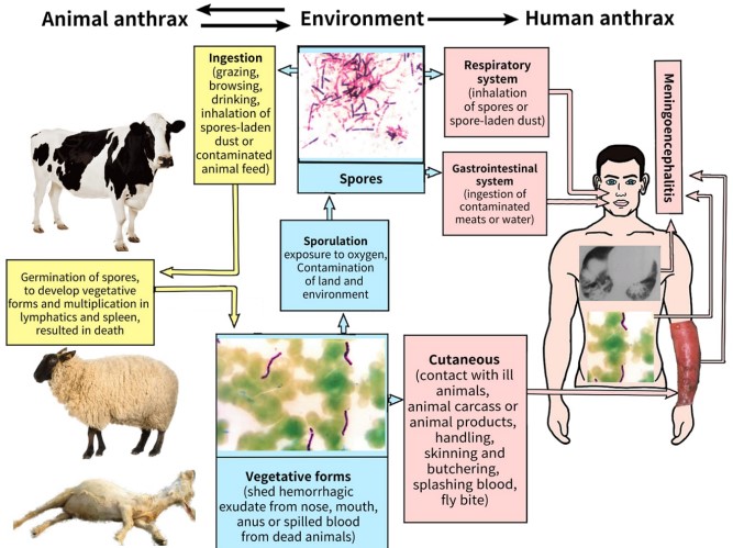 Figure 1. The life cycle of Bacillus anthracis in nature
(Source: Doganay M, et al. 2023)
The pathophysiology of anthrax is mediated by toxins secreted by Bacillus anthracis. The bacterium carries two large virulence plasmids, pXO1 and pXO2. The pXO1 plasmid encodes the protein components that make up the anthrax toxin, including edema factor (EF), lethality factor (LF), and protective antigen (PA). pXO2 plasmid encodes capsule, which helps the bacterium evade the host's innate immune response. The toxin-mediated nature of the disease is further evidenced by the fact that virulent strains of B. anthracis that lose the pXO1 plasmid become nontoxic and then become virulent when the pXO1 plasmid is reintroduced.
During infection, the vegetative form produces EF and PA, which combine to form edema toxins. The PA component binds to the cellular receptor, thereby allowing the enzyme components EF and LF to enter the cell. In the cytoplasm, LF cleaves and inactivates members of the mitogen-activated protein kinase family and NLRP1; EF rapidly increases cAMP, which activates the signaling pathway through protein kinase A. In the early stages of infection, the toxin targets cellular pathways and inhibits host innate immune responses such as neutrophil priming, chemotaxis, and chemokine production. By suppressing these critical innate immune responses at the site of infection, bacteria can escape the immune response, spread throughout the infected host, and produce large amounts of toxins.
Animal model studies have shown that both LF and EF induce vascular shock, but by different mechanisms. LF induces cytokine-independent non-hemorrhagic vascular collapse leading to hypoxic necrosis, whereas EF induces cAMP-mediated vascular dysfunction and hemorrhage. Toxins were found to cause progressive hypotension during simulated anthrax infections using a slow LF infusion model, a phenomenon associated with decreased systemic vascular resistance and left ventricular ejection fraction. These effects are also associated with progressive hepatic and renal dysfunction.
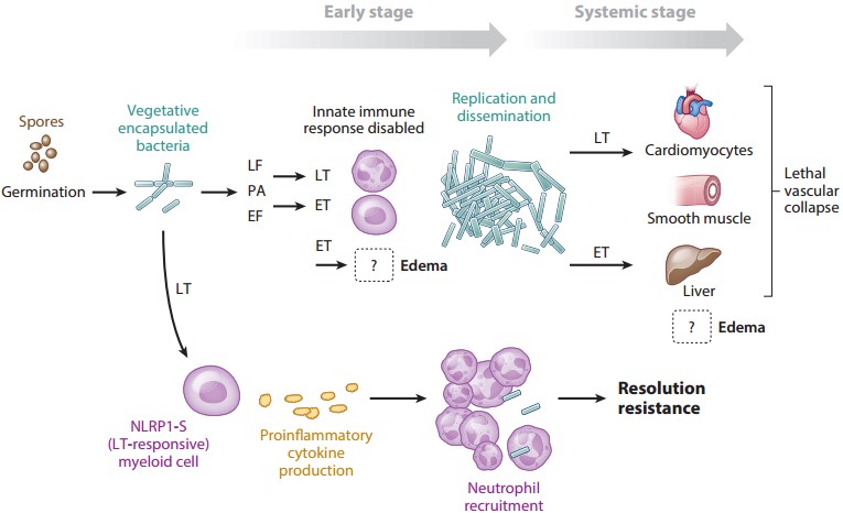 Figure 2. Spores enter the host and germinate to form capsule and toxins
(Source: Moayeri M, et al. 2015)
Cutaneous anthrax is the most common form, accounting for more than 95% of human cases, and is caused by direct contact or exposure to infected animals or contaminated animal by-products. It is usually a localized skin infection, often occurring on the face, neck, arms, or hands. The lesions begin as pruritic papules, progress to vesicles, and eventually form a typical black necrotic crust. Simple cutaneous anthrax has a mortality rate of less than 2% after treatment, but can be as high as 30% if the localized skin infection develops into systemic anthrax.
Ingestion anthrax, which is usually the result of consuming infected meat, manifests itself in two ways, as a rare oropharyngeal infection resulting in neck swelling and respiratory compromise, and as gastrointestinal anthrax, which can be accompanied by fever/chills, abdominal pain, nausea/vomiting, ascites, malaise, diarrhoea (which may be bloody) and headache. Gastrointestinal anthrax has a mortality rate of about 74%, which can be greatly reduced by early treatment after infection.
Inhalational anthrax is caused by inhalation of aerosolized spores and has historically been associated with the processing of wool, leather, or hair from infected animals. Bacillus anthracis spores can be used as a biological weapon, making inhalational anthrax a concern. The disease may have a biphasic presentation, beginning with mild symptoms of a common viral infection with fever, cough, and fatigue, followed 2-3 days later by the sudden onset of severe respiratory distress, dyspnea, and hypoxia, with a fatality rate of about 72%.
Injectable anthrax is a relatively new form of the disease that occurs only in drug users who inject drugs contaminated with Bacillus anthracis spores. Symptoms of injectable anthrax are similar to those of cutaneous anthrax, but usually occur in deeper tissues, leading to systemic disease. The mortality rate for injectable anthrax is about 25%. Meningitis occurs in 14-37% of cases and may be a primary or secondary complication depending on the route of transmission. The mortality rate exceeds 90%.
Many antibiotics target Bacillus anthracis, including but not limited to penicillin, amoxicillin, levofloxacin, and ciprofloxacin. It is worth noting, however, that anthrax is a virulence-producing disease; inhibition of bacterial growth in the host body does not result in survival, and once the toxins enter the cells in sufficient quantities, they have an effect that may last for a long time. Spores may also live for a long time in the host. There are a number of anti-PA monoclonal antibodies in clinical trials that target different stages of PA action. Some antibodies against LF and EF have also been reported to be effective. Other therapies targeting PA and its effects include receptor decoys, multivalent competitive peptides, and small molecule drugs that inhibit receptor-toxin interactions. Inhibitors that act on PA translocation function include dominant negative mutant PA proteins, small-molecule oligomerization inhibitors, β-cyclodextrin-based PA oligomer channel blockers, and other pore blockers. A different approach is to interfere with the binding of EF/LF to PA. Furin inhibitors have also been reported to have some efficacy.
The timing of anthrax treatment is very important, and early and timely treatment can significantly reduce mortality. Unfortunately, antibodies currently available for treatment after the early asymptomatic phase of infection are unable to target the toxin in the cells. Although antibody therapy can neutralize the toxin, the accumulation of the toxin between cells limits the desired effect of late or post-exposure therapy.
Almost all anthrax vaccines in use or under development use PA as the primary immunogen. Early studies showed that vaccination against PA protected against anthrax attack in a variety of animal models and that anti-PA antibody titers correlated with survival after vaccination. Live, non-encapsulated toxigenic strains have been used as vaccines, and subsequent improvements have been mainly to reduce the dose or change the route of administration. Several vaccines are currently under development, including those using less virulent and more immunogenic mutant PA variants.
A new wave of research on capsule vaccines has also led to new candidates for development. The inherent low antigenicity of capsules can be overcome by binding to a variety of carrier proteins, including PA and BSA. Capsule-conjugated vaccines conjugated to the outer membrane protein complex of Neisseria meningitidis elicited robust antibody responses but provided only partial protection in anthrax attacks, whereas immunization with capsules conjugated to peptidoglycan provided complete protection in a model of skin infection. In the development of a new method, a sortase-based conjugation approach to conjugate the capsule to domain 4 of PA resulted in a strong response to both components and provided complete protection against attack by the Ames strain in guinea pigs. Conjugation of a dominant-negative PA mutant (altered only in residues affecting the PA pore) to the capsule produced an adjuvant activity with a higher antibody response than wild-type PA conjugated to the capsule.
There are also a variety of novel oral PA vaccines, plant-based, LF-PA hybrid, and live attenuated vaccines; altered adjuvants; vaccine delivery methods; and new candidate spore and secreted protein targets under development. Despite the many projects to develop anthrax vaccines, highly immunogenic PA proteins remain the predominant and most attractive immunogens for inducing protection against this pathogen.
References
| Target | Cat. No. | Product Name | Size | Species Reactivity | Application | Detection Sample | |
| Anthrax PA | DEIASL261 | Mouse Anti-Anthrax PA83 IgG ELISA Kit | 96T | Mouse | Quantitative | Serum | Inquiry |
| B. anthracis | DEIASL269 | Rabbit Anti-Anthrax PA83 IgG ELISA Kit | 96T | Quantitative | Serum | Inquiry | |
| B. anthracis Protective Antigen | DEIABL568 | Anthrax Protective Antigen IgG Screening ELISA Kit | 96T | Qualitative | Serum, plasma | Inquiry |
| Target | Cat. No. | Product Name | Host | Isotype | Application | |
| Anthrax | CABT-51076MA | Anti-Anthrax monoclonal antibody, clone 7821 | Mouse | IgG2b | ELISA | Inquiry |
| Anthrax LF | DPAB-DC3984 | Anti-Anthrax Anthrax Lethal Factor (C-terminal) polyclonal antibody | Rabbit | IgG | ELISA | Inquiry |
| DPAB4255 | Anti-Anthrax LF (aa 47-65) polyclonal antibody | Rabbit | IgG | WB | Inquiry | |
| DPAB4234 | Anti-Anthrax Lethal Factor (CT) (C-terminal) polyclonal antibody | Rabbit | IgG | ELISA | Inquiry | |
| Anthrax PA | DPAB-DC3985 | Anti-Anthrax Anthrax PA polyclonal antibody | Rabbit | IgG | ELISA | Inquiry |
| DPAB-DC3986 | Anti-Anthrax Anthrax Protective antigen (C-terminal) polyclonal antibody | Rabbit | IgG | ELISA | Inquiry | |
| CPBT-66806RA | Rabbit Anti-Anthrax Protective Antigen Polyclonal antibody | Rabbit | IgG | WB | Inquiry | |
| DPAB4256 | Anti-Anthrax PA polyclonal antibody | Rabbit | IgG | WB | Inquiry | |
| DPAB4232 | Anti-Anthrax Protective(C-terminal) polyclonal antibody | Rabbit | IgG | ELISA | Inquiry | |
| CABT-51084MA | Anti-Protective Antigen monoclonal antibody, clone C3 | Mouse | IgG1 | ELISA, WB | Inquiry |
| Target | Cat. No. | Product Name | Expression System | Tag/Conjugate | Application | |
| B. anthracis ANTHRAX EF | CDBP5063 | B. anthracis ANTHRAX EF blocking peptide | N/A | Unconjugated | IB | Inquiry |
| B. anthracis ANTHRAX LF | CDBP5064 | B. anthracis ANTHRAX LF blocking peptide | N/A | Unconjugated | IB | Inquiry |
| CDBP5065 | B. anthracis ANTHRAX LF blocking peptide | N/A | Unconjugated | IB | Inquiry | |
| B. anthracis Protective Antigen | DAGB171 | Native Bacillus anthracis Protective Antigen PA63 [FITC] | B. anthracis | FITC | ELISA | Inquiry |
| DAGC090 | Recombinant Bacillus anthracis anthrax protective antigen | B. anthracis | Unconjugated | Inquiry |
Loading ......
