

Inquiry Basket
 Loading ......
Rheumatoid arthritis (RA) is a chronic autoimmune disease characterized by persistent synovial inflammation, progressive joint destruction, and systemic immune abnormalities. The pathogenesis of RA involves complex interactions among immune cells, pro-inflammatory cytokines, autoantibodies, and signaling pathways. Activated T cells, B cells, macrophages, and fibroblast-like synoviocytes collectively drive the inflammatory cascade, leading to cartilage degradation and bone erosion. Because immune dysregulation lies at the core of RA, effective treatment strategies aim not only to relieve symptoms but also to modulate aberrant immune responses. Methotrexate (MTX) has become the cornerstone disease-modifying antirheumatic drug (DMARD) for RA due to its well-established immunomodulatory effects, predictable clinical efficacy, and long-term safety profile when appropriately monitored.
Originally developed as an antineoplastic antifolate agent, methotrexate exerts its effects by inhibiting dihydrofolate reductase and interfering with nucleotide synthesis. However, at the low weekly doses used in RA, methotrexate functions primarily as an immunomodulatory rather than cytotoxic drug. In RA patients, methotrexate alters immune cell behavior through multiple mechanisms, including suppression of excessive immune activation, regulation of inflammatory mediators, and restoration of immune balance. These effects are cumulative and contribute to sustained disease control rather than immediate symptom relief.
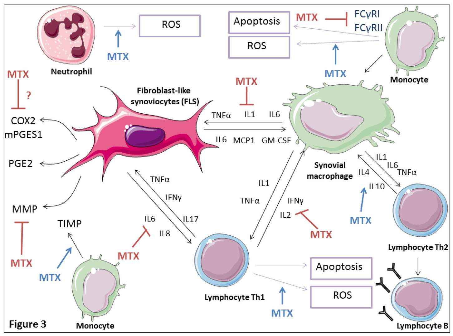 Figure 1. Immune regulatory action of low dose MTX in the RA synovial tissue.(Sources: Bedoui Y, et al.; 2019)
One of the most critical actions of methotrexate in RA is its ability to reduce the production of key pro-inflammatory cytokines such as tumor necrosis factor-α (TNF-α), interleukin-6 (IL-6), and interleukin-1β (IL-1β). These cytokines play central roles in synovial inflammation, immune cell recruitment, and joint damage. Methotrexate promotes the accumulation of extracellular adenosine, a potent endogenous anti-inflammatory mediator. Adenosine signaling inhibits cytokine release from macrophages and T cells, leading to a measurable decrease in inflammatory activity.
RA is marked by abnormal activation of CD4+ T cells and autoreactive B cells. Methotrexate reduces T-cell proliferation and shifts the immune response toward a less inflammatory phenotype. It also interferes with B-cell activation and autoantibody production, including rheumatoid factor (RF) and anti-citrullinated protein antibodies (ACPAs). These immunological changes form the biological basis for the observed reductions in disease activity scores and structural joint damage in methotrexate-treated patients.
Immune detection plays a critical role in evaluating how methotrexate treats RA at the molecular and cellular levels. Laboratory immunoassays allow clinicians and researchers to assess inflammatory markers, immune mediators, and autoantibody levels during therapy. Common immune-based indicators include cytokine profiling, RF and ACPA titers, and acute-phase reactants such as C-reactive protein (CRP). Changes in these biomarkers reflect the immunological impact of methotrexate and help determine whether immune suppression is adequate.
Enzyme-linked immunosorbent assays (ELISA), chemiluminescent immunoassays, and multiplex bead-based assays are widely used to quantify cytokines and immune biomarkers in RA patients receiving methotrexate. These techniques offer high sensitivity and specificity, making them essential tools for both clinical research and therapeutic monitoring. For example, reductions in serum IL-6 or TNF-α levels detected by immunoassays often correlate with clinical improvement and decreased synovial inflammation. Similarly, longitudinal immune detection can identify patients who respond poorly to methotrexate and may require alternative or combination therapies.
Because methotrexate suppresses immune activity, immune detection is also vital for safety assessment. Monitoring lymphocyte counts, immunoglobulin levels, and inflammatory markers helps ensure that immune suppression remains within a therapeutic range and does not predispose patients to infections or hematologic complications. Regular immune-related laboratory testing allows early identification of adverse immune effects, supporting individualized dose adjustments and long-term treatment success.
Advances in immunological detection technologies have enhanced understanding of patient-to-patient variability in methotrexate response. Immune profiling can reveal differences in cytokine signatures, immune cell activation states, and autoantibody dynamics, providing insights into treatment efficacy. As precision medicine evolves, immune detection is expected to play an even greater role in guiding methotrexate therapy, optimizing outcomes, and minimizing unnecessary immune suppression.
Methotrexate treats rheumatoid arthritis primarily by modulating immune function rather than by direct cytotoxic effects. Its ability to suppress pro-inflammatory cytokines, regulate T and B cell activity, and restore immune balance underpins its long-standing role as first-line therapy for RA. Immune detection techniques, including immunoassays and biomarker analysis, are essential for understanding methotrexate's mechanism of action, monitoring therapeutic efficacy, and ensuring immune safety. By integrating methotrexate treatment with robust immune detection strategies, clinicians and researchers can achieve more precise and effective management of rheumatoid arthritis.
Methotrexate (MTX) is a disease-modifying antirheumatic drug (DMARD) commonly used to treat rheumatoid arthritis. At low weekly doses, it modulates the immune system by reducing inflammatory cytokine production, suppressing T and B cell activation, and promoting anti-inflammatory signaling via extracellular adenosine accumulation.
Although originally developed as an antifolate chemotherapy agent, the low-dose regimen used for RA primarily affects immune cell function rather than killing cells. This allows it to control autoimmune inflammation while minimizing cytotoxic effects.
Methotrexate reduces key pro-inflammatory cytokines, including TNF-α, IL-6, and IL-1β. These cytokines drive joint inflammation and tissue damage. Methotrexate-mediated adenosine signaling inhibits cytokine release from immune cells, contributing to reduced inflammation.
Yes. Methotrexate can decrease B-cell activity and reduce the production of rheumatoid factor (RF) and anti-citrullinated protein antibodies (ACPAs), which are hallmarks of autoimmune activity in RA.
Immune detection helps monitor methotrexate's effect on the immune system. Techniques like ELISA, chemiluminescent immunoassays, and multiplex cytokine assays measure cytokines, autoantibodies, and inflammatory markers. These measurements guide therapy effectiveness and safety monitoring.
Reference
| Target | Cat. No. | Product Name | Host | Application | |
| MTX | DPATB-H81809 | Anti-Methotrexate polyclonal antibody | Goat | ELISA, RIA | Inquiry |
| DMAB-WZ0004 | Anti-Methotrexate monoclonal antibody | Mouse | IA | Inquiry | |
| DPABY-912 | Anti-Methotrexate polyclonal antibody | Sheep | ELISA, Pr* | Inquiry | |
| DPABY-070 | Anti-Methotrexate (C-terminal) polyclonal antibody | Sheep | ELISA | Inquiry | |
| DMABB-JX341 | Mouse Anti-Methotrexate monoclonal antibody, clone MTX | Mouse | ELISA, LFIA | Inquiry |
| Target | Cat. No. | Product Name | Conjugate | Application | |
| MTX | DAGB319 | Methotrexate [HRP] | HRP | IA | Inquiry |
| DAGA-351B | Methotrexate [BSA] | BSA | LFIA | Inquiry | |
| DAGA-351K | Methotrexate [KLH] | KLH | Immunogen | Inquiry | |
| DWT109 | Methotrexate Standard solution | N/A | Inquiry | ||
| DAG-WZ3635O | Methotrexate [OVA] | OVA | IA | Inquiry |
| Target | Cat. No. | Product Name | Size | Species Reactivity | Application | Detection Sample | |
| MTX | DEIA-US209 | Methotrexate ELISA kit | 96T | Human, mouse, rat | Quantitative | Serum, plasma and urine | Inquiry |
| DEIA-XYZ209 | Methotrexate ELISA kit | 96T | Human | Quantitative | Serum, Plasma | Inquiry |
Loading ......
