
Inquiry Basket
 Loading ......
Helicobacter pylori(H. pylori, Hp) was first isolated from gastric mucosal biopsy tissue in chronic gastritis and is the only microorganism found to have been isolated and cultured in the human stomach. This bacterium is spiral-shaped in vivo and rod-shaped in vitro, mainly residing in the stomach and duodenum, and is an important causative agent of human gastrointestinal diseases. Hp secretes urease, produces ammonia, can survive in strong acidic environments, and resides primarily in the stomach and duodenum. Hp mainly causes inflammatory lesions in the pyloric region of the stomach, first of all, it causes chronic gastritis, and leads to gastric ulcers and gastric atrophy, and in serious cases, it develops into gastric cancer or mucosa-associated tissue lymphoma, which is a serious danger to human health, and therefore it has been classified as a class I carcinogenic factor. The various pathologic changes caused by Hp infection result from a complex interplay of bacterial virulence, host genetics, and environmental factors, and these interactions lead to different phenotypes of chronic gastritis.
Table 1. Disease phenotypes of H. pylori infection
| Phenotype | Frequency | Localization | Effect on secretory function | Possible outcomes |
| Mild gastritis phenotype | Most patients | No specific gastric compartment predominantly affected | Normal acid secretion | Asymptomatic in most patients, no significant clinical outcome |
| Duodenal ulcer phenotype | 10-15% of patients | Antral-predominant gastritis | High gastrin and acid secretion and impaired inhibitory control of acid secretion | Dyspeptic symptoms, duodenal ulcer |
| Gastric cancer phenotype | ∽1% of patients | Corpus-predominant gastritis | Low or absent acid secretion; variable gastrin secretion | Severe atrophic gastritis and intestinal metaplasia, gastric cancer |
(Source: Malfertheiner P, et al. 2023)
Hp is one of the most important risk factors for gastric cancer, and its infection of the host activates multiple signaling pathways and proteins in the host cells, inducing epithelial-mesenchymal transition (EMT) in gastric epithelial cells, whereas aberrant regulation of EMT can lead to gastric carcinogenesis, as well as enhance the migration of tumor cells, invasion, and participate in the processes of tumor metastasis and chemoresistance. EMT was originally a normal physiologic phenomenon involved in processes such as embryonic development and adult wound healing. During EMT, the expression of many epithelial cell markers is reduced, such as ZO-1 protein, and induces the expression of mesenchymal cell markers, such as vimentin, Snail proteins, and TWIST proteins. In contrast, when EMT is abnormally regulated, epithelial cells lose polarity, become elongated in shape, and have increased cell motility and invasiveness.
 Figure 1. Progression of gastric cancer during Hp infection
(Source: Cheok YY, et al. 2021)
Cytotoxin-associated gene A (CagA) is one of the most important virulence factors of Hp. The Hp Cag pathogenicity island (CagPAI) encodes the CagA protein and a large membrane-associated transporter protein complex-type IV secretion systems (T4SSs). CagA proteins can enter gastric epithelial cells via T4SSs. In atrophic gastritis and gastric cancer, CagA-positive Hp was more pathogenic than CagA-negative Hp. In gastric epithelial cells, CagA induces EMT by down-regulating some factors or proteins, such as glycogen synthase kinase-3 (GSK-3), protein kinase CK2β, and programmed cell death protein 4 (PDCD4). CagA binds to GSK-3 and converts it to insoluble, thereby decreasing GSK-3 activity and inducing Snail-mediated EMT. CagA can also induce EMT by activating some proteins or genes, such as Yes-associated protein (YAP), aquaporin-5 (AQP5), and caudal-type homeobox protein 1 (CDX1). CDX1 is mainly involved in the transformation of gastric chronic inflammation to intestinal epithelialization promoted by CagA. It was found that in CDX1 overexpressed cells, the expression of E-cadherin was decreased, the expression of N-cadherin was increased, and the invasion and migration ability of the cells were enhanced, indicating that Hp regulates CDX1-induced EMT through CagA.
Hp secretes vacuolating cytotoxin A (VacA), an oligomeric self-transporter protein toxin that forms an anion-selective membrane channel. VacA induces intracellular production of large vacuoles from late endosomes, induces apoptosis or necrosis, induces autophagy and inhibits the proliferation of T and B cells, as well as effects on other immune cells. This cascade of actions downregulates the immune response to Hp infection and promotes host tolerance to the organism.
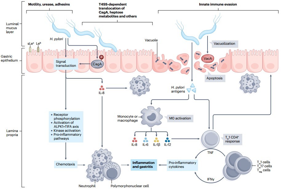 Figure 2. H. pylori infection and pathogenesis
(Source: Malfertheiner P, et al. 2023)
The discovery of Hp triggered a revolution in the treatment of digestive tract diseases, turning recurring peptic ulcers, which had been difficult to treat, into diseases that could be cured with a short course of antimicrobials. Antimicrobials are ineffective in the acidic environment of the stomach and need to target both bacteria and stomach acid. Omeprazole and amoxicillin are the classic bactericidal and antacidic duo therapy that is highly effective. Subsequent treatment for Hp eradication evolved into triple therapy, mainly bismuth or proton pump inhibitors (PPIs) plus two antimicrobials, such as a combination of PPIs, amoxicillin, and clarithromycin.
Hp on the surface of gastric mucosa is protected by the gastric mucus layer and is difficult to be removed from the stomach. Due to the long-term application of antimicrobial drugs, Hp resistance is gradually formed. In addition, when patients who have used antimicrobials are re-treated, there is an increased rate of resistance and risk of treatment failure. To address this problem, a new potassium-competitive acid blocker, vonoprazan (VPZ), an alternative triple therapy, came into use, with increased eradication of Hp, but failed to achieve significant comparative efficacy compared to standard triple therapy. In areas with high rates of clarithromycin resistance, bismuth quadruple therapy (mBCOT) consisting of bismuth, PPT, metronidazole, and tetracycline resulted in 88.2% eradication of Hp after two weeks of continuous treatment. However, the disadvantages of quadruple therapy are poor compliance and high cost. High-dose dual therapy (HDDT) consisting of a PPI and amoxicillin has better Hp eradication rates and fewer adverse effects than triple or quadruple therapy.
On the other hand, although improved drug combinations may work in the short term, efficacy decreases over time, and it may be difficult to address Hp-infectious diseases with antimicrobials alone. Therefore, changing treatment strategies is a current research topic. Newer antimicrobials can reduce side effects, improve patient compliance, and increase eradication rates. The aminoglycosides gentamicin and nertilmicin are new Hp eradicators.
The intestinal flora is disrupted by Hp infection, and the infection may be associated with metabolic syndrome in addition to causing gastroduodenal disorders. A combination of Lactobacillus rhamnosus LGG-18 and Lactobacillus salivarius Chen-08 probiotics was found to reduce gastritis and inhibit Hp-induced precancerous lesions. However, probiotic supplementation did not reduce the amount of Hp. Adjunctive probiotic therapy may aid in the restoration of the gastric microbiota after Hp eradication, but probiotic monoeradication therapy for patients with Hp infection not only has no benefit, but increases the likelihood of infection by potentially pathogenic bacteria. Vaccines are one of the new strategies to address drug resistance, and oral vaccines, which have the advantages of easy access and high compliance over injectable vaccines, can elicit humoral and cellular immune responses through the mucosa and promote the production of secretory IgA (sIgA) in the gastric mucosa.
The use of novel biomaterials as drug carriers is also a new strategy to address drug resistance, which may include biomaterials sensitive to endogenous stimuli as well as biomaterials sensitive to exogenous stimuli. A researcher has constructed a pH-responsive ROS nanomaterial consisting of metal-organic nanostructures formed by coordination of the acoustic sensitizer hematoporphyrin monomethyl ether with trivalent iron. The nanomaterials were loaded with dihydroartemisinin as a hydroperoxidant with an acidic pH-sensitive metallic shell, which was rapidly dissociated in the stomach and activated by ultrasound, where the drug produced •OH via a Fenton reaction that killed Hp.
In addition, it was found that changing the oxygen concentration had a favorable eradication effect on Hp. Some researchers have demonstrated that cultured Hp can still reproduce when the oxygen concentration drops to 4%, and that Hp does not reproduce and decreases in number over time when the oxygen concentration drops below 3%. In in vitro culture, hydrogen peroxide significantly inhibited the growth of Hp.
H. pylori infection detection methods can be categorized into invasive and non-invasive tests. Invasive tests require gastric mucosal tissue to be extracted for examination under gastroscopy, such as rapid urease test, biopsy and pathological staining examination. Non-invasive tests mainly include 13C and 14C urea breath test, Hp antibody test and fecal Hp antigen test. Compared with invasive tests, non-invasive tests are more acceptable and therefore more widely used.
The rapid urease test (RUT) is the method of choice for invasive detection of Hp infection, visualizing whether or not a patient is infected with Hp with a sensitivity of about 94% and a specificity of 95-100%. It is characterized by easy operation, rapidity, safety and accuracy for the first gastroscopy. The biopsy method (direct smear) is easy to operate and is mainly used for the rapid diagnosis of Hp, but it is easy to miss the diagnosis when the amount of bacteria is small. Histopathologic methods have a sensitivity and specificity of about 98%, and traditional Giemsa staining is costly, time-consuming, and cumbersome. Modified Giemsa staining is simpler, less time-consuming, has comparable sensitivity to traditional staining methods, and has been shown to be superior to the RUT assay. Therefore, this method can be used for routine clinical examination to provide rapid and accurate diagnosis of Hp-infected patients.
The urea breath test (UBT) has been shown to be a highly accurate test, which consists of the 13C-UBT and the 14C-UBT, both of which are similarly effective, with sensitivities and specificities of about 95%. The principle of the test is that the subject first takes 13C and 14C isotope-labeled urea, and if Hp is already present in the subject's stomach, the urease enzyme secreted by Hp will break down the urea into ammonia and 13C- and 14C-labeled CO2. By detecting the CO2 of exhaled breath, it is possible to determine whether the subject is infected with Hp or not. This method is quick and painless in diagnosis, and at the same time it can reflect the infection of the stomach, largely avoiding the limitations of biopsy specimens, and therefore it is widely used clinically for determining the infection of the patient and reviewing the patient after treatment. However, if the patient has taken an acid suppressant prior to the test, this may result in a false negative. It should be noted that 13C-UBT and 14C-UBT can cause radioactive contamination and are not suitable for pregnant women and children. Serologic antibody tests are simple and inexpensive, but they cannot distinguish whether the result is a previous or ongoing infection, so they are often used in epidemiologic investigations of infection in populations. Fecal Hp antigen is detected in feces and in blood, serum, or fingertip blood as antibody. The monoclonal antibody-based fecal antigen test has a sensitivity of 84.6% and a specificity of 94.7%. The method is simple, rapid, and easy to perform, and results are usually indicative of previous infection.
In recent years polymerase chain reaction (PCR)-based methods, such as multiplex PCR and real-time fluorescent quantitative PCR, have also been widely used for Hp detection. Molecular detection of formalin-embedded biopsy samples or RUT samples using PCR methods, of which quantitative PCR is the most appropriate, and fluorescence in situ hybridization have been shown to be highly accurate in detecting Hp, and can also be used in conjunction with molecular resistance testing.
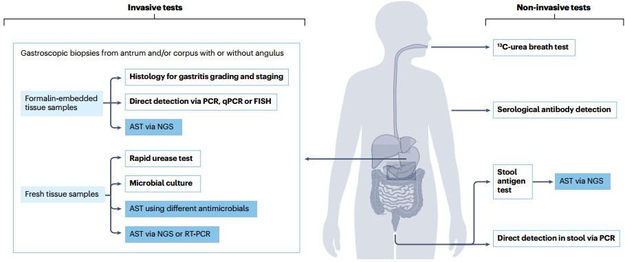 Figure 3. H. pylori diagnostic procedures
(Source: Malfertheiner P, et al. 2023)
Reference
| Target | Cat. No. | Product Name | Size | Species Reactivity | Application | Detection Sample | |
| H. pylori | DEIA341 | Human Helicobacter pylori IgG ELISA kit | 96T | Human | Quantitative and qualitative | Serum or plasma | Inquiry |
| DEIA342 | Human Helicobacter pylori IgA ELISA kit | 96T | Human | Quantitative | Serum, plasma | Inquiry | |
| DEIA343 | Human Helicobacter pylori IgM ELISA kit | 96T | Human | Quantitative | Serum, plasma | Inquiry | |
| DEIACL36 | CDSimple™ Anti-H. Pylori IgA Chemiluminescent ELISA Kit | 96T | Quantitative | Serum | Inquiry | ||
| DEIACL37 | CDSimple™ Anti-H. Pylori IgG Chemiluminescent ELISA Kit | 96T | Quantitative | Serum | Inquiry | ||
| DEIACL38 | CDSimple™ Anti-H. Pylori IgM Chemiluminescent ELISA Kit | 96T | Quantitative | Serum | Inquiry | ||
| HP-CagA-IgG | DEIA-BJ762 | Human Helicobacter pylori cytotoxinassociated gene A protein IgG ELISA kit | 96T | Quantitative | Serum, plasma, cell culture supernatants, body fluid and tissue homogenate | Inquiry |
Loading ......
