
Inquiry Basket
 Loading ......
Glucagon, produced by pancreatic α-cells, maintaining glucose homeostasis by promoting the liver to release glucose. It acting in opposition to insulin, and is crucial for ensuring stable blood glucose levels. Dysregulation of glucagon is implicated in various metabolic disorders, including type 1 and type 2 diabetes (T1D and T2D), obesity, and non-alcoholic fatty liver disease (NAFLD). In diabetes, excessive glucagon secretion contributes to hyperglycemia, making it a key biomarker for diagnosis and a potential therapeutic target.
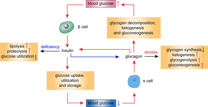 Fig. 1 Hormonal regulation of glucose homeostasis in the islet cells.
(Jia Y, et al. Front Endocrinol (Lausanne). 2022)
Blood glucose: Under normal physiological conditions, it is evident that elevated blood glucose levels suppress glucagon secretion.
Fatty acids: Longer chain fatty acids stimulate glucagon secretion through effects on cytoplasmic Ca2+ levels or fatty acid oxidation.
Amino acid: Cysteine, glycine, valine, alanine, arginine, leucine, serine, and glutamate, have been identified as stimulators of glucagon release. Additionally, high levels of leucine inhibit glucagon secretion.
Hormone: Hormonal regulators such as catecholamines (epinephrine and norepinephrine) significantly influence glucagon secretion, especially during stress or exercise.
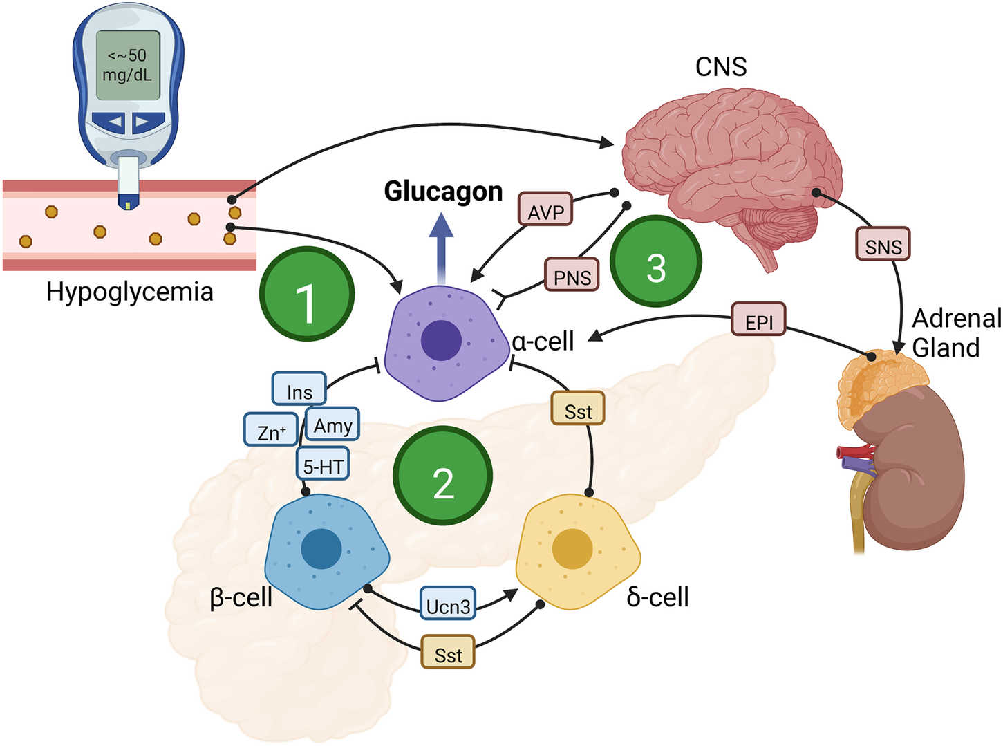 Fig. 2 Mechanisms that increase glucagon secretion in response to hypoglycemia.
(Capozzi ME, et al. Cell Metab. 2022)
Patients with T1D often exhibit inappropriate hyperglucagonemia, where α-cells fail to suppress glucagon secretion in response to elevated glucose levels. This lack of feedback inhibition exacerbates hyperglycemia by promoting hepatic glucose production, contributing to the metabolic instability observed in T1D patients. In T2D, many patients display fasting and postprandial hyperglucagonemia, despite hyperglycemia, leading to excessive hepatic glucose output. This persistent elevation of glucagon, combined with impaired insulin signaling, further drives hyperglycemia, contributing to the progressive deterioration of glucose control. Aberrations in glucagon receptor (GCGR) signaling contribute to the dysregulated glucagon secretion observed in both T1D and T2D.
Chronic hyperglucagonemia in individuals with NAFLD may contribute to hepatic steatosis by promoting lipolysis and free fatty acid flux to the liver, exacerbating lipid accumulation. Moreover, glucagon's role in stimulating hepatic glucose production may also worsen insulin resistance, creating a cycle of metabolic dysfunction that accelerates the progression of NAFLD.
Acute pancreatitis can lead to temporary dysregulation of both insulin and glucagon secretion due to pancreatic injury. In severe cases, necrotic damage to the pancreatic tissue may impair α-cell function, resulting in glucagon deficiency. This can lead to hypoglycemia, especially in patients with concomitant insulin therapy or insulin-secreting tumors (insulinomas).
Immunoassays rely on the high specificity of anti-glucagon antibodies. ELISA tests are widely used due to the high-throughput capabilities, but the accuracy may be compromised by cross-reactivity with other proglucagon-derived peptides, such as GLP-1, and the instability of glucagon during sample collection and processing. Proper sample handling, including the use of protease inhibitors and immediate freezing, is essential to mitigate degradation. RIA tests are highly sensitive but are less commonly used due to radiation safety, cost, and the need for specialized equipment. Nonetheless, RIAs can detect low glucagon concentrations with minimal cross-reactivity, making them valuable in research settings where precision is paramount.
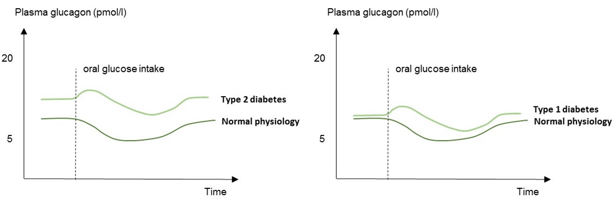 Fig. 3 Schematic illustration of plasma glucagon concentrations in patients with type 1&2 diabetes and in normal physiology (healthy subjects).
Rix I, et al. Glucagon Physiology. [Updated 2019]
Recent advancements in biosensor technology and microfluidic devices offer the potential for real-time glucagon monitoring. Biosensors integrated with antibody-based detection systems may enable continuous measurement of circulating glucagon levels, providing dynamic insights into glucose regulation and metabolic states.
Creative Diagnostics offers a wide range of glucagon and glucagon receptor antibodies validated across multiple applications, ensuring reliable and accurate results for your research and diagnostic needs.
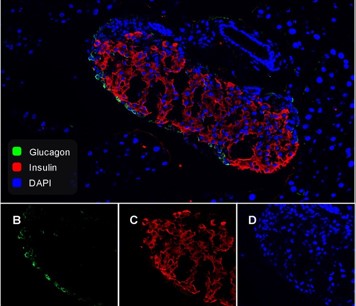
Fig. 4 Immunofluorescence analysis of frozen mouse pancreas tissue with Rabbit anti-Glucagon antibody
(Cat.No. DCABH-2679)

Fig. 5 Immunohistochemical analysis of paraffin-embedded rat pancreas tissue with Rabbit anti-Glucagon antibody
(Cat.No. DCABH-2679)

Fig. 6 Immunohistochemical analysis of paraffin-embedded Pancreas.
(Cat.No. DMABB-JX644)

Fig. 7 Binding Activity of Human GCGR with Anti-GCGR antibody.
(Cat. No. DMABB-JX647)
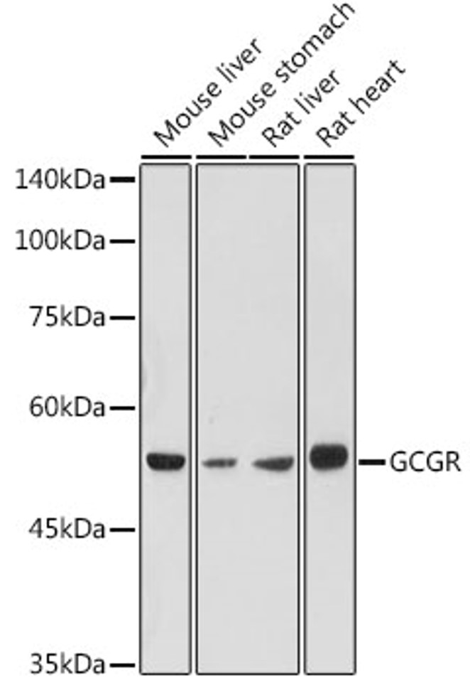
Fig. 8 Western blot analysis of extracts of various cell lines, using GCGR antibody.
(Cat. No. DPABB-JX273)
References
| Anti-Glucagon Antibodies | |||
| Cat. No. | Product Name | Application | |
| DMABB-JX644 | Mouse Anti-Human Glucagon monoclonal antibody,clone 45 | IHC, IF, ELISA | Inquiry |
| DCABY-4445 | Mouse Anti-Human Glucagon(C-term) monoclonal antibody, clone N92489 | ELISA(Cap) | Inquiry |
| DCABY-L4411U | Mouse Anti-Human Glucagon(N-term) monoclonal antibody, clone N9247 | ELISA | Inquiry |
| DCABY-L4411 | Mouse Anti-Human Glucagon(N-term) monoclonal antibody, clone N9247 [Biotin] | ELISA(Det) | Inquiry |
| DCABH-2679 | Rabbit Anti-Human Glucagon monoclonal antibody, clone KG1071 | WB, IF, IHC | Inquiry |
| DPABH-00862 | Rabbit Anti-Human Glucagon (full length) polyclonal antibody | WB | Inquiry |
| DPABH-16133 | Rabbit Anti-Human Glucagon (internal region) polyclonal antibody [Biotin] | IP, ELISA, RIA, IHC-P | Inquiry |
| CPBT-67332SP | Sheep Anti-Human Glucagon (C-Terminal) Polyclonal Antibody | RIA | Inquiry |
| CPBT-65759GH | Goat Anti-Human Glucagon (N-Terminal) Polyclonal Antibody | ELISA | Inquiry |
| DPAb2292GH | Guinea Pig Anti-Human Glucagon Polyclonal Antibody | IHC, IF, IHC-P | Inquiry |
Loading ......
