
Inquiry Basket
 Loading ......
Gastric stromal tumors (GISTs) were first proposed in 1983. They refer to a group of mesenchymal tumors with spindle cells or epithelioid cells positive for c-kit (CD117, stem cell factor receptor) staining in the gastrointestinal tract, omentum and mesentery. Stromal tumors most often occur in the stomach, with an incidence of 60% - 70%. They are gastric stromal tumors. The gross pathological findings are as follows: the tumor diameter varies from 2 to 20cm, the boundary is clear, the mass is hard, the cut surface is gray white or reddish brown, cystic or solid, and may also be accompanied by necrosis and mucus degeneration. Clinical manifestations: the ratio of male to female is 2:1, which is more common in middle-aged and elderly people. Common clinical symptoms include nausea, vomiting, epigastric pain, anemia, mass and upper gastrointestinal bleeding. Stromal tumor is a borderline tumor, which is generally divided into low-grade and high-grade malignancies.
Gist is the most common mesenchymal tumor of the digestive tract, which can occur in the stomach, small intestine, esophagus, colorectal, mesentery, liver and other parts. Immunology is mainly positive for dog1, cdll7, CD34. C-kit or PDGFRA gene gain of function mutation is an important molecular feature.

The clinical manifestations of gastrointestinal stromal tumors are diverse, and their sizes also have great differences. Because of its non-specific clinical manifestations, it is difficult to diagnose gastrointestinal stromal tumors. The discovery of gastric stromal tumor may be detected by endoscopy due to gastrointestinal symptoms, but its diagnosis and staging basically rely on CT examination. Gist has various imaging manifestations. The main imaging diagnostic points include the relationship between tumor and intestinal wall, namely, production mode, enhancement characteristics and other image features, such as calcification, necrosis, etc. The growth patterns of GIST include extraluminal type, intraluminal type, endophytic type and mixed type. Larger tumors can be accompanied by necrosis and liquefaction, and the enhancement mode is mainly obvious uneven enhancement.
| Symptoms | Descriptions |
| Multiple Population | Gastrointestinal stromal tumors account for 1-3% of gastrointestinal malignant tumors. The estimated annual incidence is about 10-20 / 1million. Most of them occur in middle-aged and elderly patients. Patients under 40 years old are rare. There is no significant difference in the incidence between men and women. 20-30% of GIST patients are malignant, and about 11-47% have metastases at the first visit, mainly in the liver and abdominal cavity. |
| Disease Symptoms | There are no specific clinical manifestations, and the course of disease can be as short as a few days to 20 years. The course of malignant GIST is relatively short, mostly within a few months. Benign or early patients are asymptomatic. The main symptoms of GIST depend on the size and location of the tumor and are usually nonspecific. Gastrointestinal bleeding is the most common symptom. Dysphagia and dysphagia symptoms are also common in gastric cardia gist. Some patients are hospitalized for ulcer perforation, which can increase the risk of peritoneal implantation and local recurrence. Ascites may occur in peritoneal dissemination, and malignant GIST may have symptoms such as weight loss and fever. |
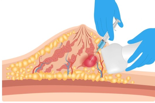
In the gross specimens, the diameter of gastrointestinal stromal tumors ranged from 1-2cm to more than 20cm, showing localized growth. Most tumors did not have a complete capsule, and occasionally a false capsule could be seen. Large tumors could be accompanied by cystic degeneration, necrosis and focal bleeding. After puncture, the tumor broke, and ulcers could also penetrate the mucosa. Most of the tumors were located in the gastrointestinal submucosa (60%), subserosal (30%) and muscular wall (10%). The boundary is clear, and those who grow into the cavity are mostly polypoid masses, often accompanied by ulceration, and grow outside the serosa to form subserosal masses. Upper gastrointestinal bleeding and touching masses are common clinical symptoms. Stromal tumors located in the abdominal cavity often have large masses. The gross shape of the tumor was nodular or lobulated, and the section was grayish white and red, uniform, hard and tough, with ulceration on the mucosal surface, bleeding, necrosis, mucilaginous change and cystic change.
1. Gastric stromal tumors occur in the body or fundus of the stomach, but rarely in the antrum.
2. Tumors tend to occur at both ends of the muscular layer of the gastric wall, and most of them grow along the vertical direction of the gastric wall. Tumors have the characteristics of large but relatively limited growth. Tumors are mostly round or quasi round soft tissue masses growing into the cavity, outside the cavity, inside and outside the cavity, and the surface can be accompanied by ulcer formation.
3. Low grade gastric stromal tumor: generally, the diameter is less than 5cm, the shape is regular, the density is uniform, punctate calcification is occasionally seen, the boundary with surrounding organs or tissues is clear, or the space occupying effect is mild, and the adjacent organs or tissues are rarely invaded.
4. Malignant stromal tumor: the diameter is more than 5cm, the growth inside and outside the cavity, the shape is irregular, the density of the mass is uneven, often accompanied by hemorrhage, necrosis, cystic change, and the boundary with surrounding organs or tissues is not clear. Gastric stromal tumors with septal enhancement are mostly malignant and have a poor prognosis, while multiple gastric stromal tumors have a poor prognosis.
5. Lymph node metastasis rarely occurs, mainly hematogenous metastasis, and there is a certain probability of planting metastasis.
The tumor was treated by surgical resection, and the main surgical method was gastric wedge resection. The operation method is suitable for tumors located in the fundus and body of the stomach, and the tumor protrudes out of the serosa, and the tumor diameter is 1.0cm ~ 3.0cm
The invasiveness of different gastrointestinal stromal tumors is indeed different. Tumor location, size, mitotic image, whether the tumor is completely resected during operation, histological type, immunohistochemical type, expression of proliferative antigen, proliferation multiple, gender, age and mutation type are related to the prognosis of the tumor, but there is no unified prognosis standard at present. Morphology and prognosis are closely related to the biological behavior of tumor and the maximum diameter and mitotic ratio of tumor. The above factors should be taken into account when evaluating the prognosis. The new classification standard combined with the location of tumor has gradually been adopted by clinicians. The location of tumor has become an independent factor for predicting the prognosis. The prognosis of stromal tumors in the stomach is better than that of stromal tumors in other parts. In addition, the difference of gene mutation sites is related to the malignant degree of tumor. Although it is not an independent prognostic factor, the recurrence rate of gastrointestinal stromal tumors with KIT exon 9 and exon 11 mutations is higher. Similar studies also show that tumor cell nuclear division count and exon 11 mutations both indicate high-risk gastrointestinal stromal tumors and poor prognosis.
| Cat. No. | Product Name | Host | Isotype | Application | |
| CABT-12875MH | Anti-KIT monoclonal antibody, clone 5G8 | Mouse | IgG | WB, IHC, sELISA, ELISA | Inquiry |
| DCABH-12110 | Anti-KIT monoclonal antibody, clone D228/480 | Mouse | IgG1 | IHC-P | Inquiry |
| DCABY-1201 | Anti-KIT monoclonal antibody, clone 677DU9.6.5 | Mouse | IgM | IF, WB | Inquiry |
| DCABH-200438 | Anti-DDX39A monoclonal antibody | Rabbit | IgG | WB, ELISA | Inquiry |
| DCABH-201591 | Anti-TLX2 monoclonal antibody | Rabbit | IgG | WB, ELISA | Inquiry |
| DMAB-DCC070 | Anti-KIT (mutation H697Y) Monoclonal Antibody, clone XH076 | Mouse | ELISA, WB | Inquiry |
| Cat. No. | Product Name | Size | Target | Species | |
| DAG-WT850B | Imatinib [BSA] | 1 mg | Imatinib | / | Inquiry |
| DAG-WT850O | Imatinib [OVA] | 1 mg | Imatinib | / | Inquiry |
| CDBP2997 | Human ANO1 blocking peptide | 50 g | TMEM16A | Human | Inquiry |
| CDBP6305 | TLX2 blocking peptide | 0.05 mg | / | / | Inquiry |
Loading ......
