
Inquiry Basket
 Loading ......
Ependymal cells, a specialized type of glial cells, line the ventricles and central canal of the brain and spinal cord. These cells possess unique characteristics that contribute to their pivotal roles in brain development, neurogenesis, and cerebrospinal fluid (CSF) production. Ependymal cells act as guardians of the neurological realm by providing physical support, regulating the exchange of substances between the CSF and brain tissue, and participating in the generation and maintenance of neural stem cells. Understanding ependymal cells and their markers is essential for studying their function, location, and potential implications for various neurological disorders.
Researchers divided ependymal cells into three subtypes based on the number and location of cilia, including multiciliated ependymal (E1 cells), ciliated ependymal (E2 cells) and uniciliated ependymal (E3 cells). Among them, E1 cells maintain normal CSF flow and participate in transport functions. E2 cells can serve as mechanical or chemical sensors of CSF flow or composition. E3 cells (β-tanycytes) are involved in regulating progenitor cell activity and sensing CSF metabolites. Another type of ependymal cells, called tanycytes, are found only in the lining of the floor of the brain's third ventricle. These cells are unique in that they have long processes and large "end feet" that connect to brain capillaries and neurons far away from the ventricles. They do not have cilia and are involved in the generation of new neurons, the generation of astrocytes, and the transport of biologically active substances between the CSF and blood vessels.
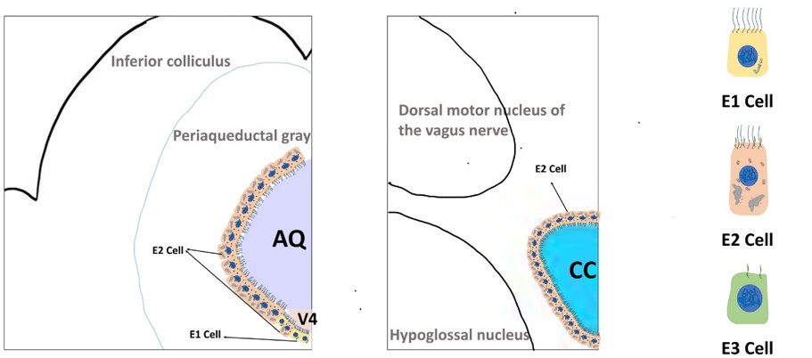 Fig. 1 Schematic diagram of the morphology and distribution of ependymal cells. (Spassky N, et al., 2005)
Accurate identification and isolation of ependymal cells are essential for studying their functions and properties. Researchers rely on specific markers to distinguish ependymal cells from other cell types in the brain. Several key markers have been identified through extensive research, providing valuable insights into ependymal cell biology and function.
Glial fibrillary acidic protein (GFAP) is a protein encoded by the human GFAP gene and plays important roles in cell morphology maintenance, cell mitosis, and cell interconnections. GFAP is expressed by multiple cell types in the central nervous system (CNS), including astrocytes and ependymal cells during development. Immunohistochemical labeling experiments indicate that ependymoma cells, whether neoplastic cells or normal cells, may display positive immunolabeling for GFAP, vimentin, and cytokeratin.
Forkhead box protein J1 (FoxJ1) is a transcription factor primarily associated with the development and function of ciliated cells. Ependymal cells possess motile cilia on their apical surfaces, which are vital for the movement of CSF and circulation within the ventricles. Diseases associated with FOXJ1 include Ciliary Dyskinesia and Primary Ciliary Dyskinesia. In addition, acetylated α-tubulin (TUBA1A), a ciliary protein, is also considered a reliable marker of ependymal cells.
To distinguish tanycytes from ependymal cells, researchers used the Allen Brain Atlas to identify genes expressed only in ependymal cells. By this approach, Rarres2 was determined to be selectively expressed in ependymal cells and serve as a candidate reliable ependymal marker.
Additional markers of ependymal cells, identified by RNA-sequencing, include TMEM212, CCDC153, TM4SF1, and MIA.
At Creative Diagnostics, we offer a wide range of high-quality antibodies and reagents for ependymal cell research. Our products are meticulously designed and validated to ensure accurate and reliable results in your experiments. Whether you are studying ependymal cell function, localization, or their role in disease, our innovative solutions can support your research endeavors. Contact us today to explore our comprehensive portfolio of ependymal cell markers and take your research to new heights.
References
Loading ......
