
Inquiry Basket
 Loading ......
CL is a mitochondrial type of phospholipid, so named because it was firstly isolated from the heart. Cardiolipin has two phosphate molecules and three glycerol molecules attached to four fatty acid molecules. Since phosphate groups are deprotonated by the pH environment, cardiolipin is usually negative. Cardiolipin is mostly located in the inner mitochondrial wall, and it makes up a quarter of all phospholipids. And in the outer mitochondrial membrane too, which makes up about 4% of it, especially around interfaces between the inner and outer membranes. Here cardiolipin is able to get to the outer mitochondrial wall and the mitochondrial tip to the cytoplasm.
The majority of the lipids are generated in the ER and sent to the mitochondria, although cardiolipin and phosphatidylethanolamine also produce in the mitochondrial inner membrane. Cardiolipin is made by a reaction that involves precursors (G-3-P), CTP and enzymes (GPTA). The process can generally be divided into three steps:
1. Formation of PA and its transport from the mitochondrial outer membrane to the inner membrane:
PA is produced through two successive acylation reactions of G-3-P. Mitochondrial PA is located near ER membranes associated with mitochondria and must traverse the outer membrane, intermembrane space, and inner membrane to reach the inner leaflet of the inner membrane for cardiolipin synthesis.
2. Conversion of PA to cardiolipin on the matrix-facing side of the inner membrane. This involves several reactions:
First, PA reacts with CTP under the catalysis of CDS to produce CDP-DAG and pyrophosphate. Next, CDP-DAG reacts with G-3-P, catalyzed by Pgs1, forming PGP. PGP is then dephosphorylated by a phosphatidylglycerophosphatase enzyme to generate PG.
3. Remodeling of cardiolipin: In the final step, PG combines with CDP-DAG under the action of cardiolipin synthase to produce cardiolipin.
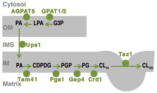 Figure 1. Cardiolipin biosynthesis in mitochondria
(Source: Schlame M, et al. 2017)
Three translocations transport CL from the inner leaflet of the inner membrane to the surface of the mitochondria:
1. Inner leaflet outer leaflet of inner membrane.
2. From outer leaflet of the inner membrane to inner leaflet of the outer membrane.
3. From inner leaflet to outer leaflet of outer membrane.
Cardiolipin is essential for mitochondrial integrity. Because cardiolipin causes negative curvature elastic stress in lipid bilayers, it’s also involved in keeping mitochondrial inner membrane cristae curvy. Cardiolipin can also attach to proteins on the mitochondrial inner membrane like cytochrome c to help bind the membrane. For example, cardiolipin deficiency has been shown to interfere with ATP synthase’s oligomeric construction, directly affecting the formation of the cristae in the inner membrane of the mitochondria.
Cardiolipin can also interact with complexes within the electron transport chain, linking oxidative phosphorylation complexes together. Complex III and Complex IV can go into a supercomplex, for instance, transporting proton and electrons to make ATP. In the process of apoptosis, cardiolipin will be redistributed – e.g., it becomes more abundant in the outer leaflet of the inner mitochondrial membrane and on the cell surface. Furthermore, apoptotic stimulation can also drive cardiolipin metabolic turnover, which decreases its mitochondria. In apoptosis, cardiolipin is also a signaling integrator that coordinates cytochrome c, caspase-8 and the pro-apoptotic protein Bid.
 Figure 2. The role of CL in mitochondrial metabolism and as a signaling platform
(Source: Dudek J, et al. 2019)
aPL are autoantibodies fused to phospholipids or phospholipid-binding proteins. It’s all anticardiolipin antibodies, lupus blood thinners and anti-beta-2-glycoprotein 1 (beta-2-GP1) antibodies. ACAs are negative cardiolipin-targeted autoantibodies on platelets and vessel endothelial cells, a connective tissue serving as a middleman between the immune system and the blood coagulation machine. Antiphospholipid syndrome (APS) – an autoimmune condition common to aPL and classified clinically as chronic arterial and venous thrombosis, frequent miscarriage, systemic lupus erythematosus. These findings in the lab in APS usually suggest persistent medium to high titers of aPL in the bloodstream. This is the pathological mechanism of cardiovascular involvement in APS through endothelial dysfunction and activation of the complement system through aPL. That can result in carotid intima-media thickening, carotid plaque growth and ankle-brachial index increase.
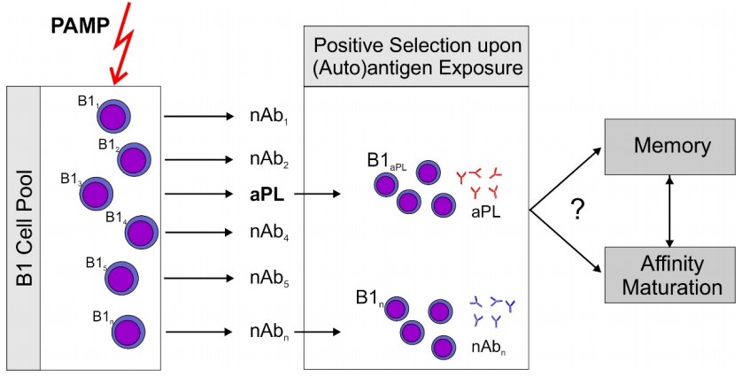 Figure 3. Proposed sequence of events leading to aPL
(Source: Lackner KJ, et al. 2016)
OAPS is the term we hear a lot about antiphospholipid syndrome in pregnancy when we have continued miscarriage, early embryo death, preterm birth and placental death. Anticardiolipin antibodies moderate to severe were linked to preeclampsia. Preeclampsia patients have more antiphospholipid antibodies than normal volunteers. And the anticardiolipin and anti-beta-2-GP1 antibodies tend to be higher in early stage severe preeclampsia, so there’s a correlation.
ACA can lead to RSA by various pathological processes. ACA interacts with negatively charged phospholipids on the membrane of vascular endothelial cells and shuts off beta-2GP1’s anticoagulant function. ACA-beta-2GP1 modulates TLR4/MyD88/NF-κB pathway and upregulates adhesion molecules, tissue factor, IL-6 and intracellular adhesion molecules in endothelial cells. This promotes platelet aggregation and thrombus formation. Additionally, ACA binding to endothelial phospholipids damages vascular endothelial cells. Its destroyed cells produce arachidonic acid, which blocks prostacyclin production and swells the prostacyclin/thromboxane ratio. A swollen endothelium also causes platelets to stick and bundle up and release thromboxane A2, thereby narrowing placental blood vessels. Thrombosis of the placental veins causes decreased placental blood flow and, therefore, a reduction in blood flow to the placenta, leading to a miscarriage or stillbirth. ACA also decreases the levels of progesterone and beta-HCG in the endometrium, making the embryo imposable. Research has revealed that RSA patients who have positive ACA show increased levels of pro-apoptotic cytokines (IL-1, IL-6, TNF-α). Mutating the TLR4/MyD88/NF-κB pathway might suppress tissue factor (TF), vascular cell adhesion molecule-1 (VCAM-1), and related proteins and thus prevent them.
 Figure 4. Summary of the proposed pathogenesis of aPL–mediated clinical problems
(Source: Garcia D, et al. 2018)
We know from several studies that ACA is highly related to ischemic stroke. ACA-positive individuals who had cerebral infarction were more likely than their ACA-negative counterparts to develop multiple lesions; ACA serum concentrations increased in association with lesion number. A clinical follow-up study revealed that ACA-positive stroke patients had a significantly higher recurrence rate compared to the control group. When the IgG titer of ACA exceeded 40 GPL, the probability of recurrent thrombotic events (43.3%) was significantly higher than when it was below 40 GPL (7.7%), with the difference being statistically significant. Researchers suggest that higher serum concentrations of ACA are associated with an increased recurrence rate. It could be because of some of the other characteristics we have seen in ACA-positive stroke patients: more and larger infarct lesions, NIHSS scores at onset which reflect worsening conditions, and multiple risk factors for cerebrovascular disease.
ACA is associated with ischemic stroke risk factors: hypertension, diabetes, dyslipidemia, anticoagulant-fibrinolytic system dysfunction, smoking. Increased blood pressure damages vascular endothelial cells, exposing membrane phospholipids as autoantigens and leading to production of ACA. The presence of high blood glucose can lead to ACA production through inflammatory cytokines release and hence endothelial dysfunction. ACA also worsens vascular injury by inhibiting the production of prostacyclin (PGI2) and activating platelet aggregation that leads to thrombosis and diabetes complications. ACA also interferes with lipid metabolism, causing atherosclerosis to become more rapid and creates thrombosis. The strong positive relationship was discovered with triglycerides and a weak negative relationship with HDL and apolipoprotein A-I.
References
| Target | Cat. No. | Product Name | Size | Species | Application | Detection Sample | |
| Cardiolipin | DEIABL326 | Cardiolipin Screen ELISA Kit | 96T | Quantitative, Qualitative | Serum | Inquiry | |
| DEIA-BJ895 | Human Anti-Cardiolipin Antibody IgA ELISA kit | 96T | Human | Quantitative | Plasma, Serum | Inquiry | |
| DEIA1708 | Human Cardiolipin IgG ELISA Kit | 96T | Human | Quantitative | Human serum or plasma | Inquiry | |
| DEIA1709 | Human Cardiolipin IgM ELISA Kit | 96T | Human | Semi-quantitative | Human sera | Inquiry | |
| DEIA1710 | Human Cardiolipin IgA ELISA Kit | 96T | Human | Semi-quantitative | Human sera | Inquiry | |
| DEIA1812 | Human Anti-Cardiolipin IgG/IgM ELISA Kit | 96T | Human | Quantitative | Serum, plasma | Inquiry | |
| DEIA1813 | Human Anti-Cardiolipin IgG/IgM ELISA Kit | 96T | Human | Quantitative | Serum, plasma | Inquiry | |
| DEIA2360 | Cardiolipin IgG/IgA/IgM ELISA Kit | 96T | Human | Qualitative | Serum | Inquiry | |
| DEIA-Z89013 | Human Cardiolipin (aCL) IgM ELISA Kit | 96T | Human | Quantitative | Serum, plasma | Inquiry | |
| DEIA1708M | Human Cardiolipin IgM ELISA Kit | 96T | Human | Quantitative and Qualitative | Human serum or plasma (EDTA, citrate, heparin). | Inquiry | |
| DEIA6134 | Cardiolipin IgM ELISA Kit | 96T | N/A | Quantitative | Saliva | Inquiry | |
| CRLS1 | DEIA-FN328 | Human CRLS1 (Cardiolipin synthase) ELISA Kit | 96T | Quantitative | Serum, plasma, cell culture supernatants, tissue homogenate | Inquiry |
| Target | Cat. No. | Product Name | Expression System | Tag/Conjugate | Application | |
| Cardiolipin | CABT-L4631 | Human Anti-Cardiolipin polyclonal antibody | Human | IgG | ELISA | Inquiry |
| DMABB-JX223 | Human Anti-Human cardiolipin monoclonal antibody, clone D24A | Human | IgA | ELISA | Inquiry | |
| CABT-L6468Z | Human Anti-cardiolipin monoclonal antibody, clone D24G | Human | IgG | ELISA | Inquiry | |
| CABT-L6469Z | Human Anti-cardiolipin monoclonal antibody, clone D24M | Human | IgG | ELISA | Inquiry | |
| DPAB-CL019 | Anti-Cardiolipin polyclonal antibody | Human | IgG | ELISA | Inquiry |
| Target | Cat. No. | Product Name | Host | Isotype | Application | |
| Cardiolipin | DAG-WT3257 | Purified Cardiolipin | Bovine heart | N/A | Standard | Inquiry |
| DAG-WT830 | Cardiolipin [GST] | N/A | GST | Immunoassays | Inquiry |
Loading ......
