
Inquiry Basket
 Loading ......
Baculovirus: A double-stranded, circular, and supercoiled DNA virus. They possess between 80 and 180 kb genome containing between 90 and 180 genes. Baculoviruses have been discovered in many species of invertebrates, but they infect and spread mostly through arthropods in the wild. Upon entering the cell nucleus, the virus particles are bound into a virogenic stroma (VS) compartment, situated in the centre of the infected cell's nucleus, where viral DNA is organized and replicated. Although the genomes of baculoviruses are so different, there is one gene set shared by all of the sequenced baculoviral genomes. They are called core genes and a handful of the proteins they code for are the structure of the virus particles, used to build and transfer the nucleocapsid, to make nucleic acids and to inoculate host gut tissues.
During the long process of co-evolution with insect hosts, baculoviruses have developed a unique two-phase life cycle, resulting in two morphologically distinct viral particle phenotypes: occlusion-derived viruses (ODVs) and budded viruses (BVs). ODVs are primarily responsible for oral infections within the intestines of live insects, while BVs mainly mediate cell-to-cell infections. In the early stages of viral infection, the nucleocapsids assembled in the host cell nucleus are transported to the cytoplasm and then bud off into the extracellular environment to form BVs, leading to the spread of the virus between cells or tissues. In the later stages of infection, mature nucleocapsids acquire membrane structures from the inner nuclear membrane to form ODVs, which are subsequently embedded in polyhedral proteins to create occlusion bodies (OBs). These OBs are released into the environment, facilitating the transmission of the virus among hosts and contributing to viral epidemics.
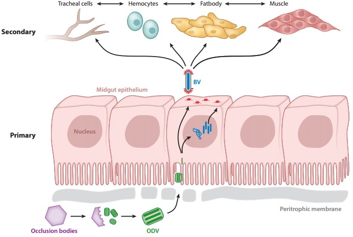 Figure 1. Baculovirus virion phenotypes and their roles in the infection of host tissues
(Source: Blissard GW, et al. 2018)
The assembly of viral nucleocapsids generally involves the formation of a protein shell and the encapsidation of the viral genome. Although there are significant differences in the assembly mechanisms of DNA viruses, most of them typically employ two main strategies for nucleocapsid assembly. The first strategy involves the direct binding of the viral genome to free capsid proteins or subunits, which encapsulates the genome as the capsid structure forms. This mechanism is commonly observed in small DNA and RNA viruses. The second assembly mechanism separates the formation of the protein shell from the encapsidation of the viral genome. In this case, an empty capsid is first formed, and then molecular motors are used to concentrate and package the viral genome into the capsid, a process primarily seen in viruses with linear genomes. Baculoviruses utilize this second assembly mechanism. In host cells infected by baculoviruses, a viral replication structure (VS) can form within the nucleus, consisting of a fibrous electron-dense matrix and an electron-transparent inner matrix space. The fibrous electron-dense matrix serves as the area for baculovirus DNA replication, and as DNA replication progresses, capsid proteins are transported to the VS region to assemble into capsid structures. The nucleocapsid structure comprises three parts: a cap structure, a cylindrical shell, and a basal structure. Research indicates that the encapsidation of baculovirus genomic DNA occurs within pre-assembled capsids, a process believed to initiate at the cap structure.
Baculoviruses, after genetic engineering modifications, can serve as powerful vectors with significant applications in biopharmaceuticals and gene therapy. The BEV system has been successfully utilized for the production of large quantities of recombinant proteins. Developed in the last century, the BEV system initially achieved recombinant viruses by replacing the polyhedrin gene of AcMNPV. Subsequently, various methods for constructing recombinant viruses have been developed and optimized to enhance the yield and quality of target proteins. However, compared to other expression systems, there is still considerable room for improvement in the BEV system. Techniques such as baculovirus genetic engineering and the introduction of genes for complex mammalian N-glycosylation can be employed to enhance protein stability and expression levels.
Scientists initially constructed recombinant baculoviruses using homologous recombination methods with viral genomes and plasmid vectors. However, the complexity of multiple blue-white screening processes and the extremely low recombination rates of the viruses necessitated further optimization of this method. A research team successfully utilized replication-defective baculovirus particles to undergo homologous recombination with a vector carrying the target gene. Once recombination was successful, the baculovirus regained its replication capability, enabling rapid preparation of recombinant baculoviruses and efficient screening of secreted recombinant proteins, including those that are difficult to express. After baculovirus infects insect cells, it expresses proteins in a sequential manner. The very late promoters polh and p10 exhibit strong protein expression capabilities. When using these very late promoters to express proteins, the cells are already in the late stages of viral infection, which diminishes their ability to produce proteins effectively, leading to cellular pathology and reduced protein stability. To address this issue, researchers have combined different promoters with polh or p10 to enhance protein expression levels in the BEV system. For instance, using both vp39 and polh promoters together facilitates the expression of foreign proteins. Additionally, through bioinformatics analysis of the baculovirus genome, researchers have attempted to knock out certain non-essential viral genes that negatively impact recombinant protein production and stability. For example, knocking down the vcath and chiA genes—responsible for infecting larval cells and causing their lysis—has been shown to adversely affect recombinant protein expression. The deletion of these genes can improve the stability and yield of secreted recombinant proteins.
The IC-BEVS is one of the four major expression systems in genetic engineering. The baculovirus expression system has a well-developed mechanism for post-translational modifications, allowing expressed proteins to form complex tertiary or quaternary structures. This enables foreign proteins to closely resemble the structure of natural proteins, resulting in high protein activity and immunogenicity comparable to that of natural proteins. Its promoters can efficiently drive the high expression of target genes, with the expression levels of target proteins reaching approximately 50% of the total cellular protein. The complete genome of the baculovirus contains multiple natural strong promoters, allowing for the insertion of relatively large fragments of foreign genes and enabling the simultaneous expression of multiple genes. Co-expression of several genes can overcome the weakness in immunogenicity associated with single-component subunit vaccines, and virus-like particles (VLPs) can also act as adjuvants. Additionally, the insect cells used, such as Sf21, Sf9, or High-Five, can be cultured in adherent or suspension conditions at 27°C without carbon dioxide, making the operation relatively simple and providing a natural advantage for industrial production. Although the baculovirus genome can be transduced into mammalian cells, it cannot replicate and does not possess the characteristics of viral proliferation and intercellular dissemination. It also does not stimulate a strong immune response in mammals or cause functional damage to them, but it is capable of transferring foreign genetic material into mammalian cells.
When expressing foreign genes using the baculovirus expression system, there are generally three expression strategies: (1) Construct recombinant baculoviruses that control the foreign gene under eukaryotic or viral promoters, such as the polyhedrin promoter and the P10 promoter, to infect insect cells and achieve target protein expression. (2) Fuse the foreign gene with the baculovirus envelope protein GP64 gene for displaying the target protein on the surface of the viral envelope. (3) Fuse the foreign gene with an occlusion body localization sequence to anchor the target protein in a specific area.
 Figure 2. Baculovirus engineering
(Source: Mansouri M, et al. 2018)
Despite this, several factors limit the large-scale application of the BEVS in practice. For instance, the high cost of cell culture media and serum on the market poses a significant challenge, as expressing target proteins requires substantial amounts of insect cell culture media and serum, leading to increased production costs. Additionally, recombinant viruses infecting insect cells can induce the expression of genes such as viral or cellular caspases, triggering apoptosis and resulting in a shorter cell lifespan, which negatively impacts the yield of target protein expression. Furthermore, there are notable differences in N-glycosylation modifications between insect cells and mammalian cells, resulting in altered structure and function of humanized glycoproteins expressed in insect cells. Consequently, target proteins are more susceptible to degradation by proteases present in insect cells.
At present, baculovirus vector system is used in the development of animal virus vaccines, such as foot-and-mouth disease virus, sheep pox virus, porcine epidemic diarrhea virus and pseudorabies virus. Relevant proteins have been expressed in the baculovirus system and are associated with an extreme immune reaction in the host. However, most vaccines remain limited to laboratory studies. On the other hand, baculovirus DNA has been successfully engineered as a vector for expressing foreign proteins, achieving successful expression of proteins such as human IFN-β, human growth factor, human 2,6-sialyltransferase, human granulocyte-macrophage colony-stimulating factor, and human anti-albumin immunoglobulin G1. Vaccines derived from the BEVS have been brought to market and are used for the prevention and treatment of cervical cancer, prostate cancer, lipoprotein lipase deficiency genetic disorders, influenza, and more.
The IC-BEVS can also be utilized for the production of enveloped and non-enveloped VLPs. This system offers several advantages in VLP production, such as higher protein expression yields compared to bacterial or yeast systems, the presence of complex post-translational modification pathways, and the ability to form multi-protein VLPs. However, it also has significant drawbacks; the N-glycosylation pattern of glycoproteins expressed in insect cells is simpler than that in mammalian cells, and the simultaneous production of enveloped baculoviruses and VLPs increases both the cost and complexity of purification. If improvements can be made to its glycosylation patterns, the IC-BEVS may become the most suitable system for producing VLP vaccines.
The baculovirus-mediated gene transfer system has gained widespread attention in gene therapy. In this system, baculovirus vectors are introduced into mammalian cells through transduction, allowing the therapeutic gene expression cassette controlled by mammalian promoters to function within the cells. For instance, researchers have utilized baculovirus vectors to deliver the glutamine synthetase gene, finding that this gene was expressed in MA104 epithelial cells, L6 myoblasts, and myotubes. This model experiment lays the groundwork for future clinical treatments for patients with human glutamine synthetase deficiency.
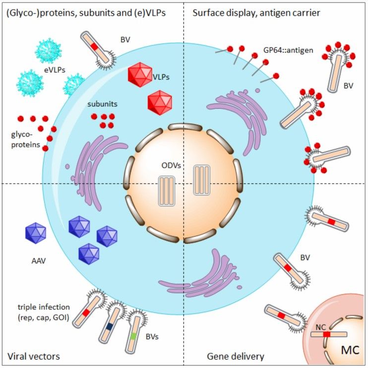 Figure 3. Overview of the various applications of the baculovirus expression system
(Source: van Oers MM, et al. 2015)
References
| Target | Cat. No. | Product Name | Host | Isotype | Application | |
| Baculovirus gp64 | DMABT-Z60787 | Anti- baculovirus gp64 Monoclonal antibody, Clone CeX7 | Mouse | IgG2b | FC, IHC, IP, WB | Inquiry |
| DPAB-CS24001 | Rabbit Anti-Baculovirus Envelope gp64 Polyclonal antibody | Rabbit | IgG | ELISA | Inquiry | |
| DMAB-CS24020 | Mouse Anti-Baculovirus Envelope gp64 Monoclonal antibody, clone BdW6 | Mouse | IgG2b | WB | Inquiry | |
| DMAB-CS24021 | Mouse Anti-Baculovirus Envelope gp64 Monoclonal antibody, clone N112 | Mouse | IgG1 | Neut | Inquiry | |
| Baculovirus p35 | DPAB-CS24002 | Rabbit Anti-Baculovirus p35 Polyclonal antibody | Rabbit | IgG | WB, ICC, IF, IHC | Inquiry |
| Baculovirus | DMAB9358 | Anti-Baculovirus Monoclonal antibody | Mouse | IgA | IP, WB, IHC | Inquiry |
| CABT-CS074 | Mouse Anti-Baculovirus Envelope gp64 protein Monoclonal antibody, clone BdW2 | Mouse | IgG2a | FC | Inquiry |
Loading ......
