Astrocytoma refers to a tumor formed by astrocytes. According to literature reports, astrocytomas account for 13% to 26% of intracranial tumors and 21.2% to 51.6% of gliomas, with more males than females. Astrocytic tumors can occur in any part of the central nervous system, generally more common in the cerebral hemispheres and subthalamic regions in adults, and more common in children under the tentorium. Those on the screen are more common in the frontal and temporal lobes, followed by the parietal lobe, and the least common in the occipital lobe. It can also be seen in the optic nerve, thalamus, and the third ventricle; Those below the screen are mostly located in the cerebellar hemisphere and fourth ventricle, and can also be seen in the vermis and brainstem of the cerebellum. Astrocytoma is an invasive growth tumor, and most tumors have the possibility of recurrence after resection. After recurrence, the tumor can evolve into anaplastic astrocytoma or glioblastoma multiforme.
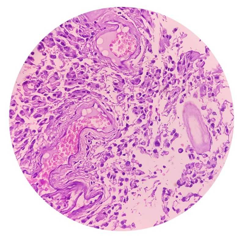 Figure 1. Astrocytoma.
| Etiology | Details |
| Astrocytoma | Tumors are mainly located in the white matter and grow infiltratively. Solid tumors have no obvious boundaries and are mostly not limited to one lobe of the brain. Tumors that grow outward can invade the cortex and damage deep structures inward. They can also cross the midline of the corpus callosum and invade the contralateral hemisphere. Tumors with cystic changes can be called "cystic tumors". Astrocytoma located in the cerebellum is often a large cyst with tumor nodules on the cyst wall. The cyst wall is composed of fibrous connective tissue and glial fibers. Therefore, only removing the tumor nodules can achieve the goal of curative treatment of the tumor. |
| Interstitial or malignant astrocytoma | Mainly found in the brain, the tumor is large and sometimes invades several lobes or crosses the midline to invade the contralateral hemisphere. The tumor tissue is gray red in color and has a soft texture. It grows infiltratively in the brain and has a certain boundary with the surrounding brain tissue. Tumor cells can infiltrate and grow into the cortex, forming a "satellite phenomenon" around neurons. There are cystic changes and small focal hemorrhagic necrotic lesions. |
| Hair cell type astrocytoma | Hair cell type astrocytoma grows slowly and originates from neuroepithelial tissue tumors. Tumors are more likely to occur in the white matter of the midline structure and the cerebellar hemisphere, with the most typical occurrence occurring in the funnel region, sometimes referred to as funnel tumors; Gliomas that occur in the optic nerve are called optic nerve gliomas. Tumors that occur in the anterior visual pathway, hypothalamus, and brainstem have unclear boundaries, are mostly substantial, and have abundant blood supply. |
| Clinical Manifestations | Details |
| Astrocytoma | Growth is slow, the course of the disease often lasts for several years, with an average of 3.5 years. Most patients show slow progressive development, and epilepsy is often the first symptom. 50% of patients have epilepsy onset, and most patients have headaches, psychomotor muscle weakness, vomiting, and obvious consciousness disorders. Most patients with neurological examination have optic disc edema and neurological disorders. Nearly half of the patients experience limb weakness, while a small number of patients experience speech difficulties, sensory disorders, and changes in vision. |
| Anaplastic astrocytoma | The duration of the disease is on average 6-24 months shorter than that of astrocytomas. The main clinical symptoms of cerebral hemisphere lesions are headache, psychiatric symptoms, limb weakness, vomiting, speech difficulties, visual changes, and drowsiness. Neurological examination can reveal symptoms of hemiplegia, optic disc edema, neurological damage, hemianopia, and hemiparesthesia. The onset of the disease is progressively worsening, and some may experience sudden deterioration. Early intracranial pressure increases can be present in brain tumors, including hemiplegia, neurological weakness, memory loss, confusion, epilepsy, and endocrine disorders. |
| Hairy cell astrocytoma | Generally, the course of the disease is long. For tumors located in the orbit, the main manifestation is visual impairment accompanied by painless exophthalmos. There may be different types of hemianopia, strabismus, and optic nerve atrophy. For tumors located in the optic chiasm, bilateral vision is often affected, including optic disc edema, strabismus, optic nerve atrophy, and headache. Hypothalamic tumors often have endocrine disorders, diencephalic syndrome, and precocious puberty. Tumors with a diameter of 2cm or more can cause hydrocephalus. |
1. Neuroelectrophysiological examination
Electroencephalography (EEG) can be helpful for patients with epilepsy as the initial symptom, mainly manifested as focal low amplitude slow waves, and some may show widespread moderate or severe abnormalities. Visual evoked potential (VEP) examination is helpful for optic gliomas and temporal occipital lobe tumors, while brainstem auditory evoked potential (BAEP) is helpful for the diagnosis of tumors in the brainstem, cerebellum, and other areas.
2. X-ray examination
Most patients show increased intracranial pressure on X-ray plain films of the skull. Partial calcification can be seen in the form of dots or arcs. Optic nerve tumors can show enlargement of the optic foramen and cause deformation of the anterior clinoid process and sellar tubercle.
3. CT examination
Fibrous and protoplasmic astrocytomas often present as low-density, uniform and consistent CT images, with no significant space occupying effect. There are no hemorrhagic or necrotic lesions within the tumor, and no obvious edema around the tumor. Except for a few cases, injection of contrast agents generally does not enhance or slightly enhances. On CT, anaplastic astrocytoma presents as a low-density or heterogeneous mixture of low-density and high-density lesions.
4. MRI examination
Astrocytoma shows low signal on T1W and high signal on T2W on MRI. MRI can clearly display the degree of tumor infiltration into brain tissue. After enhancement, astrocytomas generally do not show enhancement, and a few tumors may have peripheral patchy mild enhancement shadows. Benign astrocytomas exhibit low signal intensity on T1 weighted images and high signal intensity on T2 weighted images, with uniform signal intensity and mild peritumoral edema.
Diagnosis can generally be made based on the patient's clinical manifestations and auxiliary examinations. Lumbar puncture should be considered contraindicated for patients with significant intracranial hypertension. Generally, astrocytomas often present with varying degrees of increased intracranial pressure. Cerebrospinal fluid examination shows that most white blood cells are normal while protein content is increased. This is particularly evident when the tumor is close to the ventricles or subarachnoid space, but normal cerebrospinal fluid protein content cannot rule out the presence of the tumor.
At present, it is widely advocated that surgery should be pursued as much as possible for astrocytomas, and postoperative adjuvant radiotherapy and/or chemotherapy should be used as appropriate; For those who cannot undergo surgery, stereotactic radiotherapy and/or chemotherapy can be used.
1. For small and well-defined solid low-grade gliomas located in deep brain and/or important functional areas (such as brainstem and thalamus), SRT treatment alone can be used to reduce radiation damage to normal brain tissue.
2. For high-grade gliomas with poor radiation sensitivity (such as anaplastic astrocytomas, glioblastomas, etc.), regular conventional radiation therapy can be used after surgery, and SRS/SRT can be used as a supplement or booster therapy to increase the radiation dose to tumor tissue and improve local control rate.
3. For low/high-grade gliomas that recur after surgery and radiotherapy and have a small volume, simple treatment with SRS/SRT can be considered.
| Cat. No. | Product Name | Host | Isotype | Application | |
| DMABT-H21795 | Anti-Astrocytomas monoclonal antibody, clone K2-42 | Mouse | IgM | WB, ICC, IHC-P | Inquiry |
| DMAB5390MH | Anti-Ubiquitin monoclonal antibody, clone GQN2 | Mouse | IgG1 | IHC | Inquiry |
| DMAB5405MH | Anti-NCAM1 monoclonal antibody, clone 124D4.E6 | Mouse | IgG1 | IHC | Inquiry |
| Cat. No. | Product Name | Size | Target | Species | |
| CDBP5810 | NRN1 blocking peptide | 0.05 mg | Inquiry | ||
| CDBP1692 | Human KLF16 blocking peptide | 100 g | KLF16 / DRRF | Human | Inquiry |
| CDBP1861 | Human MELK blocking peptide | 100 g | MELK | Human | Inquiry |