


Inquiry Basket
 Loading ......
Aspartic proteases are a large, evolutionarily and functionally diverse family of proteolytic enzymes. These enzymes have two highly conserved aspartic acid residues that function in a collaborative manner in the catalytic cleavage of peptide bonds. Aspartic proteases have been identified in all types of organisms ranging from retroviruses to fungi, plants, and mammals. They perform unique, often specific physiological roles for the organisms in which they are present. Aspartic proteases have been implicated in several human diseases. The plasmepsins of the malaria parasite, the HIV-1 protease, and the secreted aspartic proteases from Candida albicans and related species are attractive drug targets, being the main targets for antimalarial drug development, a key enzyme in HIV replication whose inhibition is an important therapeutic strategy against AIDS, and a major virulence factor, respectively.
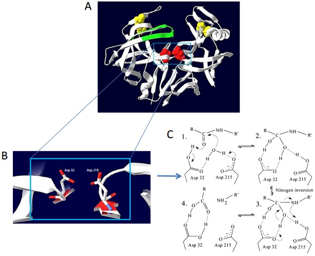 Figure 1. Three-dimensional structure and mechanism of action of a typical aspartic protease. (Source: Theron LW, et al. 2014)
Table 1. Examples of Common Aspartic Proteases
| Source Category | Enzyme Name | Primary Source | Key Functions and Characteristics |
| Mammals | Pepsin | Gastric Chief Cells | Key enzyme for dietary protein digestion; activated and functions in a highly acidic environment. |
| Renin | Kidney | Rate-limiting enzyme of the renin-angiotensin system; regulates blood pressure and fluid balance by cleaving angiotensinogen. | |
| Cathepsin D | Lysosomes (ubiquitous in cells) | Major enzyme for intracellular protein degradation and turnover; dysfunction is associated with neurodegenerative diseases. | |
| β-secretase 1 (BACE1) | Neurons and other cells | Cleaves amyloid precursor protein (APP); the rate-limiting step in generating Aβ peptide, a key pathogenic factor in Alzheimer's disease. | |
| Fungi/Microbes | Chymosin (Rennet) | Calf Stomach (traditional); Microbial Fermentation (e.g., Mucor, Aspergillus) | Key coagulant in cheese making; specifically cleaves κ-casein in milk, causing it to curdle. |
| Secreted Aspartic Proteases (SAPs) | Candida albicans | Critical virulence factors; involved in fungal adhesion, invasion of host tissues, and immune evasion. | |
| Plants | Cardosins / Cynarases | Artichoke (Cynara cardunculus) | Traditionally used in Southern European cheese production as a plant-based alternative to rennet. |
| Viruses | HIV-1 Protease | Human Immunodeficiency Virus Type 1 (HIV-1) | Essential for viral replication; cleaves viral polyprotein precursors into mature, functional proteins. A primary target for anti-AIDS drugs. |
| Parasites | Plasmepsins | Plasmodium falciparum | Degrades host hemoglobin in the parasite's digestive vacuole to provide amino acids for the malaria parasite; a potential target for antimalarial drug development. |

Aimee DPABH-23467
Great
Sweileh DCABH-10545
Works Great!
Al-Sehaibany DCABH-9425
Great product
Shattock DCABH-3141
Function good
The Anti-CTSD monoclonal antibody can be utilized in WB
Yes, the buffer of Anti-TMED10 monoclonal antibody, clone FQS0148(C) contains 0.01% Sodium Azide.
No, the Anti-PSEN2 monoclonal antibody, clone BQT 37 is unconjugate.
The buffer used in Anti-BACE2 monoclonal antibody is included In 1x PBS, pH 7.4.
catalysis. At catalytic pH optimum one of the Asp residues is deprotonated (negatively charged carboxylate form) and the other is protonated (electrically neutral carboxylic acid form). The negatively charged Asp residue acts as a general base and the other as a general acid.
Water Molecule Activation and Nucleophilic Attack: The catalytic process begins with a water molecule that is precisely bound between the two Asp residues. The deprotonated Asp residue, acting as the general base, abstracts a proton from this water molecule, thereby "activating" it into a more potent nucleophile—a hydroxyl ion. This hydroxyl ion then launches a nucleophilic attack on the carbon atom of the carbonyl group in the scissile peptide bond of the substrate.
Formation and Stabilization of the Tetrahedral Intermediate The nucleophilic attack results in an unstable, high-energy tetrahedral intermediate. The oxyanion of the intermediate is stabilized by hydrogen bonding with surrounding amino acid residues including the protonated Asp residue serving as general acid.
Peptide Bond Cleavage and Product Release: The protonated Asp residue serves as the general acid and donates its proton to the nitrogen atom of the scissile peptide bond. This step results in cleavage of the carbon-nitrogen bond. The newly created C-terminal and N-terminal products are then released in sequence from the active site, and the enzyme is returned to its original state.
Many aspartic proteases, such as HIV-1 protease and BACE1, possess a flexible β-hairpin loop structure known as a "flap," which overhangs the active site cleft. The flap is in an open conformation in the absence of a substrate. The flap closes down when a substrate or inhibitor binds. The substrate is enclosed in the active site and bulk water molecules are excluded. The active site provides a catalytic microenvironment.
 Figure 2. Mechanism of amide bond hydrolysis by aspartic proteases. (Source: Eder J, et al. 2007)
Alzheimer's disease is the most prevalent neurodegenerative disorder. There are two key pathological hallmarks that are well-known to underlie AD: extracellular amyloid-β (Aβ) plaques and intracellular neurofibrillary tangles (NFTs). For several decades, the "amyloid cascade hypothesis" has been the basis for AD pathogenesis, whereby Aβ production and deposition is believed to lead to a series of downstream events (Tau pathology, neuroinflammation and neuronal death). However, already at the beginning of this cascade two aspartic proteases play a critical role.
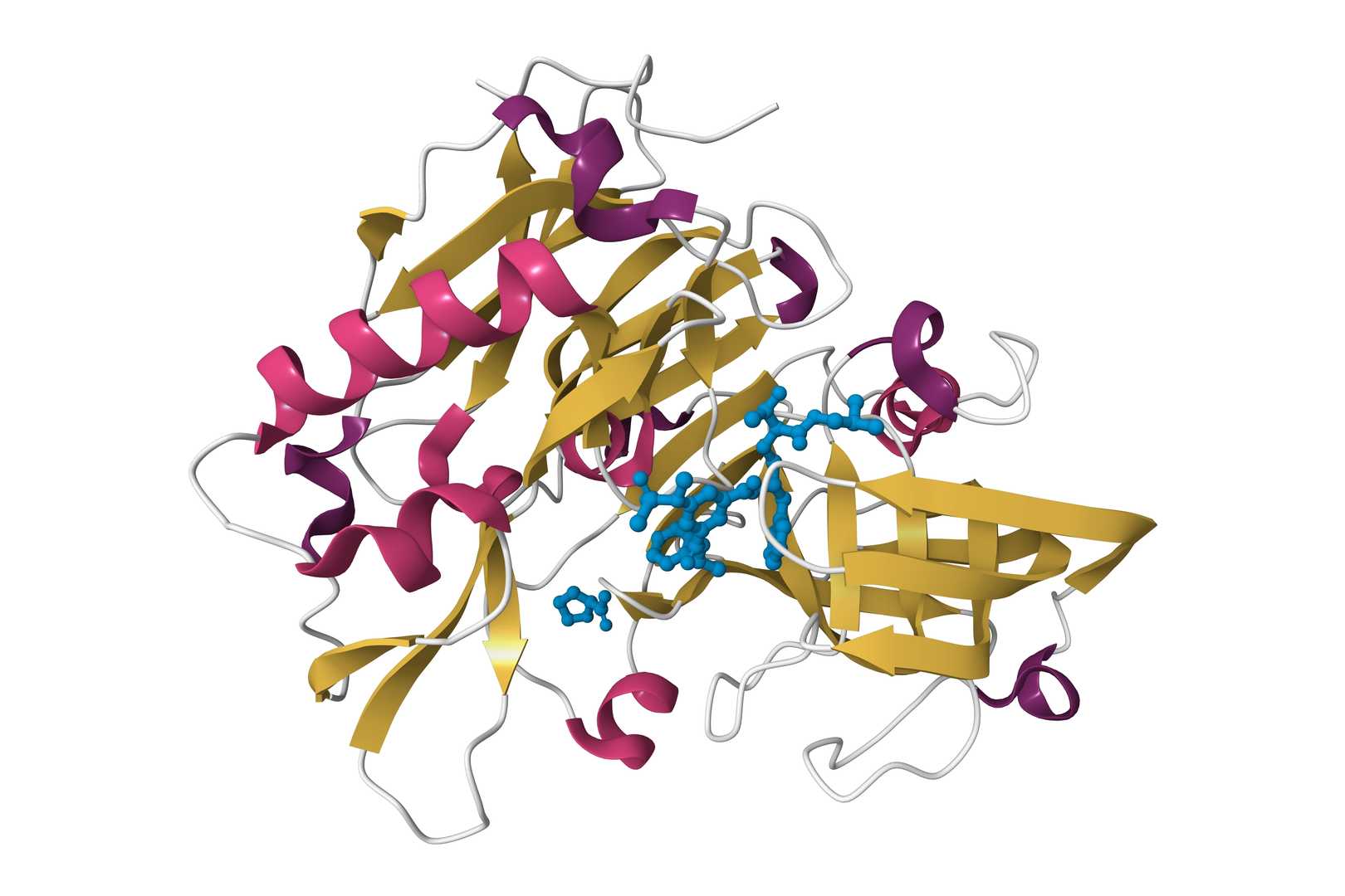
BACE1:
The Rate-Limiting Step in Aβ Production
Amyloid precursor protein (APP) is a type I transmembrane protein that is ubiquitously expressed. APP can be cleaved by α-secretase in the Aβ sequence during normal physiological conditions, which is referred to as the non-amyloidogenic pathway. However, under disease conditions, APP is processed through the amyloidogenic pathway. BACE1 is responsible for the first and rate-limiting step of this pathway. The discovery of BACE1 is a landmark in AD research since it unambiguously identified an aspartic protease that is required for Aβ production as a β-secretase. BACE1 cleaves APP just outside the N-terminus of the Aβ region to produce a membrane-bound fragment (C99) that includes the full-length Aβ sequence.
γ-Secretase:
The final release of Aβ
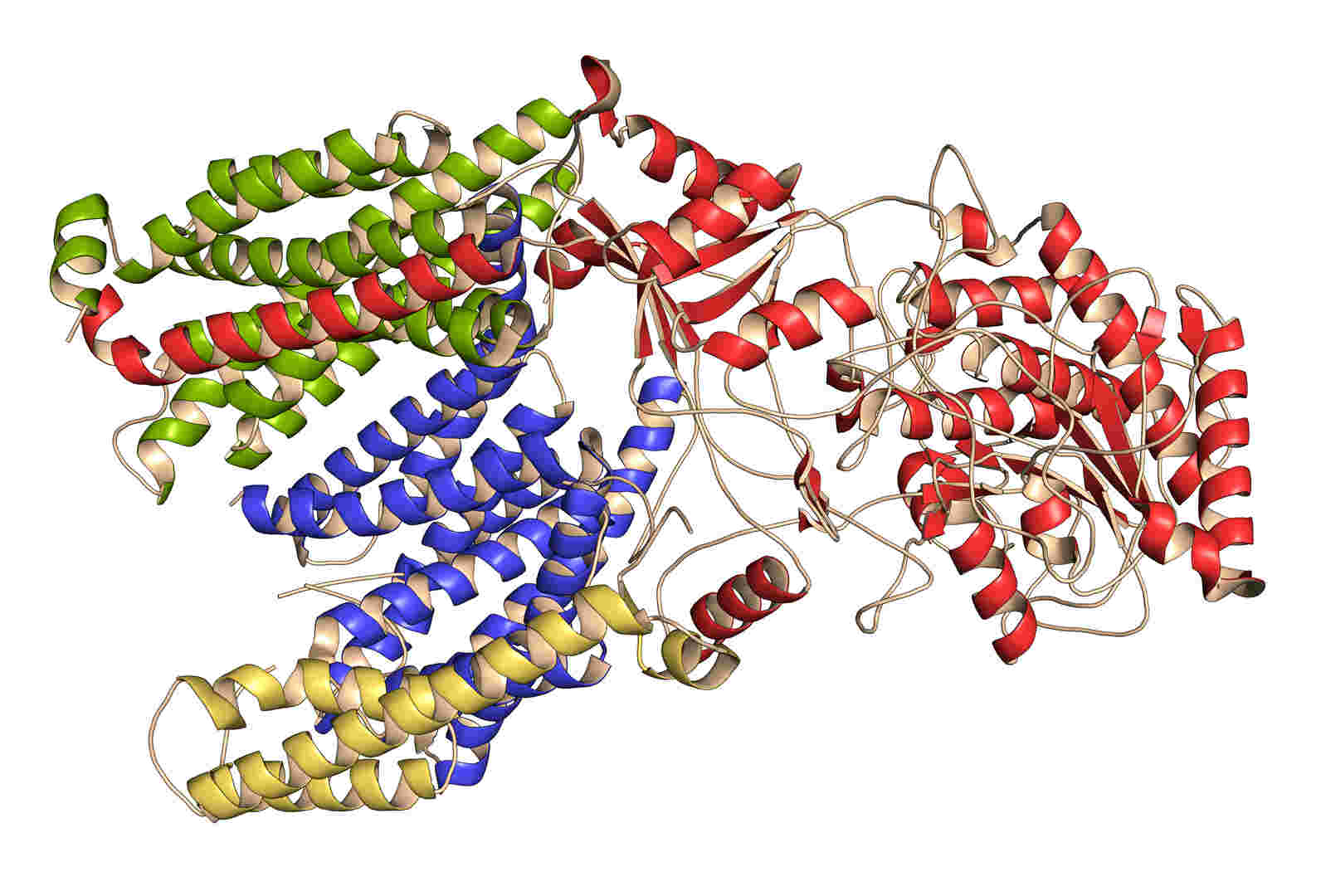
The C99 fragment is further processed by the γ-secretase complex. γ-Secretase is an atypical multi-subunit intramembrane protease complex with Presenilin (a rare aspartic protease) as the catalytic component in its core. The primary activity of γ-secretase takes place in the lipid bilayer. γ-Secretase cleaves the transmembrane domain of C99, liberating the Aβ peptide from the membrane. The cut is imprecise, resulting in Aβ peptides of different lengths. Aβ42 subtype (42 amino acids long), which is the most aggregation-prone and neurotoxic form of the peptide, is considered to be the main factor in the formation of amyloid plaques.
References
Loading ......
