
Inquiry Basket
 Loading ......
Programmed cell death, also known as apoptosis, is a highly regulated process that plays crucial roles in various physiological and pathological processes, including development, tissue homeostasis, immune response, and cancer. Understanding the molecular mechanisms underlying apoptosis is essential for developing effective therapeutic strategies for diseases involving aberrant cell death, such as cancer and neurodegenerative disorders.
Apoptosis is initiated by a complex signaling cascade that involves a diverse array of molecules and pathways. One of the key events in apoptosis is the activation of caspases, which are a family of cysteine proteases that cleave a wide range of cellular substrates, leading to the characteristic morphological and biochemical changes associated with apoptosis. Caspases are activated through two main pathways: the intrinsic and the extrinsic pathway.
The intrinsic pathway is regulated by members of the Bcl-2 family of proteins, which regulate mitochondrial outer membrane permeabilization (MOMP). MOMP results in the release of pro-apoptotic factors, including cytochrome C, from the mitochondrial intermembrane space into the cytosol. Cytochrome C then binds to the adaptor protein apoptotic protease-activating factor 1 (APAF-1), leading to the formation of the apoptosome complex and the activation of caspase-9, which in turn activates the effector caspases-3 and -7.
The extrinsic pathway is initiated by the binding of extracellular death ligands, such as tumor necrosis factor (TNF) and Fas ligand (FasL), to their corresponding death receptors on the cell surface. This results in the recruitment of adaptor proteins, such as the Fas-associated death domain (FADD), and the activation of caspase-8, which then activates caspases-3, -6, and -7.
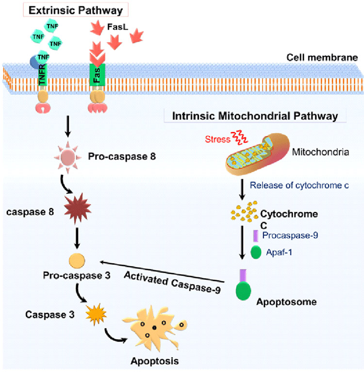 Fig. 1 Schematic representation of apoptosis mechanisms. (Bhosale P B,et al., 2020)
| Early-Stage Events |
|
| Mid-Stage Events |
|
| Late-Stage Events |
|
Annexin V assays are used to detect the early stages of apoptosis. Phosphatidylserine, a membrane phospholipid, is normally located in the inner membrane leaflet of viable cells. However, during apoptosis, phosphatidylserine is translocated to the exposed membrane surface, serving as a signal for phagocytic cells to attack. Annexin V is a protein that can bind specifically to cellular membranes and has a high affinity for phosphatidylserine-containing membranes. Annexin V conjugated with a fluorescent dye can be used to detect phosphatidylserine exposure on the cell surface by flow cytometry or fluorescence microscopy.
When the cells in the early stage of apoptosis have not changed significantly under the microscope, the mitochondrial membrane potential in the cell has already begun to change, including the increase in mitochondrial membrane permeability and the decrease in transmembrane potential. The decrease in membrane potential is considered to be the earliest biological change in apoptosis. If the membrane potential is destroyed, apoptosis will be irreversible. Some lipophilic cationic fluorescent dyes, such as rhodamine 123 and JC-1 can be tightly combined with the mitochondrial matrix, and the degree of decrease in mitochondrial membrane potential is positively correlated with the decrease in mitochondrial dye aggregate ability.
Caspase-3 is considered the key enzyme for various factors leading to apoptosis. Under physiological conditions, it exists in the form of a zymogen. Its activation indicates the beginning of cell execution of the apoptosis program, and it is a sign that apoptosis enters an irreversible stage. At present, the activation of caspase-3 has not been found in necrotic cells. Therefore, whether cells are in apoptosis or necrosis can be judged by detecting caspase-3 activity. There are two kinds of detection content and detection activity. The detection content is Caspase quantification or relative quantification by Western blot, immunohistochemistry, or immunofluorescence. The detection of activity involves the use of fluorescein-coupled short peptides that can be specifically recognized by Caspase. After Caspase cuts short peptides, fluorescein will be released, and Caspase activity can be determined based on fluorescence intensity.
DNA fragmentation is a hallmark of the late stages of apoptosis. The DNA break formed by apoptosis will expose the 3'-OH end of the DNA chain, and the terminal deoxynucleotidyl transferase (TdT) can catalyze the nucleic acid polymerization reaction of the 3'-OH end of the broken DNA. The addition of dUTP labeled with fluorescein, peroxidase, alkaline phosphatase, or biotin allows specific detection of apoptotic cells in situ after color reaction. TUNEL assays provide biologically important data on DNA damage and late-stage apoptosis.
We offer a range of apoptosis assay kits that are designed for the detection of various apoptosis markers. For example, the Annexin V-FITC Apoptosis Detection Kit can be used to detect early apoptosis by flow cytometry or fluorescence microscopy. The Caspase-3 Colorimetric Assay Kit measures caspase-3 activity in cell lysates or tissue samples using a substrate that changes color upon cleavage. The Mitochondrial Membrane Potential Assay Kit provides a simple and reliable method for measuring changes in mitochondrial membrane potential using the JC-1 dye. The DNA Fragmentation Detection Kit uses the TUNEL assay to detect DNA fragmentation in apoptotic cells.
Our high-quality apoptosis assay kits and tools can help researchers investigate different aspects of apoptosis and facilitate the development of effective therapies for diseases involving dysregulated cell death.
Reference
Loading ......
