
Inquiry Basket
 Loading ......
ALP is a well-known osteoblastic marker, clinically, a loss-of-function mutation in the liver/bone/kidney ALP (ALPL) gene has been linked to a severe skeletal deformity disease termed hypophosphatasia (HPP) that is characterized by bone mass loss and, consequently, pathological fractures. ALPL in humans and mice strongly suggests that ALPL is necessary for postnatal bone formation and that the bone deformities are related to the degree of ALPL deficiency. ALP has been widely used as a diagnostic index to evaluate bone formation capacity in osteoporosis.
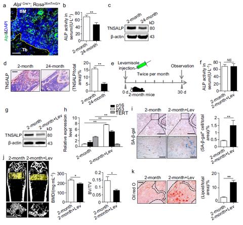 Fig1. Declined expression of Alpl in the bone marrow is associated with bone ageing characteristics.
(Source: Bone Research, June 2018.)
Bone ageing is often manifested as a progressive decrease in bone mass and a parallel increase in marrow fat as shown by a microcomputer tomography (μCT) analysis and Oil Red O staining. Alpl is expressed in many organs and tissues, especially in the kidney, liver and bone, as shown in AlplCre/+; Rosa26mTmG/+ mice. Using two ageing mouse models, scientist revealed that bone ageing characteristics could be associated with a decreased Alpl expression in the BM.
Bone ageing, which is the main risk factor for primary osteoporosis, results in a decrease in bone mass and a parallel increase in marrow fat. At the cellular level, bone marrow (BM) mesenchymal stem cells (MSCs), which are common progenitors of osteoblasts (OBs) and adipocytes in the BM, undergo senescence along with bone ageing. MSCs and osteoblastic cell lineages have been shown to spontaneously release ATP, which plays a central role in bone physiology. Tissue nonspecific ALP (TNSALP), which is encoded by ALPL, is used as a surface marker for the prospective isolation of MSCs. TNSALP appears to be involved in the metabolism of nucleotides and can sequentially hydrolyze ATP, ADP and AMP, the TNSALP level has been reported to be inversely correlated with the extracellular ATP concentration in neurocyte culture medium. Therefore, there is a hypothesize that Alpl may regulate ATP homeostasis in MSCs and subsequently lead to the fate switch in MSC differentiation and senescence.
The AMP-activated protein kinase α (AMPKα) pathway is directly regulated by the ATP level, AMPKα pathway also plays a role in bone physiology. The Inhibition of the AMPKα pathway in MSCs promotes differentiation at the expense of osteogenic differentiation. Alpl deficiency results in an excessively high level of extracellular ATP due to enhanced ATP release and reduced ATP hydrolysis, which is subsequently internalized by MSCs, increasing the intracellular ATP level. This elevation inhibits the AMPKα pathway and contributes to the cell fate switch of MSCs. To summarize, Alpl orchestrates lineage differentiation and senescence in MSCs through ATP-mediated regulation of AMPKα, thereby playing an important role in bone ageing.
Osteoclast Immunology Reagents
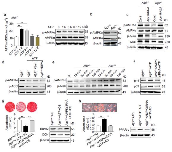 Fig. 2 ATP-mediated AMPKα pathway inactivation contributes to MSC dysfunction.
(Source: Bone Research, June 2018.)
bone, Alpl is localized on the entire cell surface of pre-osteoblasts and has long been used as an osteoblastic marker. MSCs are common progenitors of OBs and adipocytes in BM and abundantly express Alpl, which is consistent with our results. However, the mechanism by which Alpl regulates the function of MSCs remains elusive. Alpl is expressed in one-cell stage embryos and has been proposed to ensure that ES cells are maintained in an undifferentiated state, suggesting that Alpl plays one or several functions in these undifferentiated cells and during their multipotential differentiation.
Loading ......
