
Inquiry Basket
 Loading ......
AAT is a single chain glycoprotein belonging to the Serpin. It's the largest protease inhibitor in human plasma, produced and excreted mainly by the liver. The AAT can also be produced by monocytes, macrophages and epithelial cells. AAT plasma levels are in normal subjects at 1.5 to 3.5 g/L, but as an acute phase reactant it rises in response to inflammation.
AAT's active site, methionine at position 358, makes the enzyme specific. AAT can also block the proteases neutrophil elastase (NE), cathepsin G and proteinase 3 (PR3), which prevents the breakdown of tissue. Among these interactions, AAT has the highest rate constant for binding with NE, indicating a stronger specificity for this protease.
AAT gets modified post-translationally by glycosylation on the endoplasmic reticulum (ER). Three of the asparagine residues at 70,107 and 271 on the polypeptide chain are changed in the ER by adding N-glycosidically linked oligosaccharides processed in the ER and Golgi apparatus. AAT glycosylation also changed with disease conditions, and AAT glycosylation correlated with AAT activity and abundance in diseases ranging from inflammation to cancer.
 Figure 1. Molecular model of glycosylated alpha-1 antitrypsin
(Source: O'Brien ME, et al. 2022)
AAT is an inhibitor of proteases, so it serves to balance proteases and antiproteases. AAT shields connective tissues of the lungs from serine proteases such as neutrophil elastase (NE). This protective role is strongest in genetic diseases where AAT deficiency is present, and in which a patient is much more likely to develop emphysema young in life. When AAT is not sufficient, or there is a functional mutation in AAT, NE will over-digest elastin, severing the elastic qualities of lung tissue and causing respiratory issues like adult-onset chronic obstructive pulmonary disease (COPD). In the normal situation, AAT is produced in the liver and released into the blood. However, defective AAT cannot enter the circulation and accumulates in the liver, potentially resulting in cirrhosis in both adults and children. Like all serine protease inhibitors, AAT features characteristic secondary structures, including β-sheets and α-helices. Mutations in these regions can lead to the formation of non-functional protein aggregates that accumulate in the liver, causing infantile cirrhosis.
Circulating AAT can be internalized by endothelial cells through a clathrin-mediated process, where it exerts its non-classical functions by inhibiting TNF-α, caspase-3, and intracellular cysteine proteases. This action reduces the levels of apoptosis and mitigates tissue damage, providing protective effects within the cells. The immunofluorescence showed that AAT co-localises with caspase-3 in apoptotic cells — something it doesn't do in living cells. It's PR3, a proteinase 3 found in the cytoplasm of adult neutrophils, that triggers caspase-3, setting neutrophils on fire. AAT also anti-apoptoticly affects other cell types, including pancreatic -cells and vascular endothelial cells, but no one is sure exactly how.
Table 1. Known binding partners to alpha-1 antitrypsin in health and disease
| Binding Partner | Disease Processes | |
| Plasma and circulating cells | Apolipoprotein B-100 | Atheroma |
| IgA complexes | Rheumatoid arthritis, myeloma | |
| Fibrinogen | Healthy | |
| IgK light chains | Myeloma | |
| HSP 70 | Diabetes mellitus | |
| Complement C3 | Complement activation | |
| Serum albumin | Not specified | |
| IL-8 | Emphysema/pneumonia | |
| Intracellular | Heme | Not specified |
| Caspase 3 | Apoptosis, emphysema | |
| Caspase 1 | Apoptosis | |
| Extracellular/Tissue | IgA complexes | Synovial fluid/rheumatoid arthritis |
| Aggrecanase 1 | Synovial tissue/OA | |
| Bile acids | Bile | |
| NO | Inflammation/innate immunity |
(Source: O'Brien ME, et al. 2022)
AATD is a metabolic disease that results from point mutations, insertions and deletions of the AAT gene. Mutant forms of alpha-1 antitrypsin bunch and even lump in liver cell endoplasmic reticulum, so AAT cannot enter the bloodstream. Such liver-stored mutant proteins can cause liver damage and even cirrhosis. Lung tissue, however, has an equilibrium protease-antiprotease milieu at all times. If AAT is impaired, this harmony is badly disturbed, causing injury to lung tissue and eventually pathology.
AATD is an autosomal recessive genetic liver metabolic disease in which genes for the SERPINA1 gene are not functioning properly. Genetic defects in the SERPINA1 gene disrupt AAT release and fold the protein together in the liver to drive liver damage, cirrhosis and even liver cancer. Furthermore, AAT deficiency causes elastic tissue to be destroyed in the lungs leading to pneumonia and emphysema.
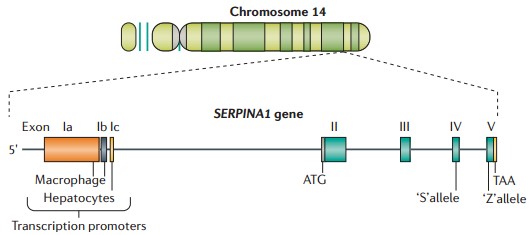 Figure 2. SERPINA1 and α1-antitrypsin
(Source: Greene CM, et al. 2016)
AAT gene is polymorphic — more than 150 allelic variants have been detected to date. They come in four forms: (1) Normal (normal plasma AAT concentration and function); (2) Deficient (low plasma AAT concentration); (3) Null (invisible plasma AAT); (4) Dysfunctional (normal plasma AAT concentration but abnormal function). There are mainly two types of AAT mutations present in AATD patients, the S (moderately low protein level) and the Z (severely low protein level) at rates of roughly 0.02 and 0.05 respectively.
Classic AATD is caused by homozygous Z mutations (Glu342Lys), accounting for approximately 95% of clinical AATD cases. The Z mutation leads to abnormal folding of the AAT protein, with only 10% to 15% correctly folding and being secreted as active monomers into the extracellular space. Most Z mutant AAT proteins don't fold in the right way, aggregating in the endoplasmic reticulum of liver cells. At the clinical level, it usually leads to neonatal hepatitis, cirrhosis and liver cancer in newborns and adults. For people with the Z mutation, a single nucleotide change takes place in exon V of the AAT gene; Glu342 is replaced by Lys. This dissociates the once stable salt bond between Glu342 and Lys290, and the hydrogen bond between Glu342 and Thr203, thus modifying the stability of the AAT molecule.
AATD can be diagnosed early enough for patients to adopt preventive measures and receive appropriate treatment, but there is no perfect diagnostic tool yet. Laboratory tests mainly include plasma level assessments, protein phenotyping, genetic testing, and histopathological examinations. The first step in diagnosing AATD typically involves measuring serum AAT levels. During inflammatory responses, levels of C-reactive protein (CRP) and AAT may both increase, but since CRP is more sensitive than AAT, it is recommended to test for CRP simultaneously. If CRP results are abnormal, AAT test results may not be considered valid. On the other hand, if CRP is normal and AAT is below normal, type of mutation can be further identified by protein phenotyping, PCR or direct sequencing. In patients with liver disease, histopathological analysis of the liver tissue can check liver function and exclude causes of liver disease. Babies with persistent jaundice or nonspecific liver dysfunction should be tested for AATD. In adults with chronic respiratory diseases, incompletely reversible asthma, bronchiectasis, primary ciliary dyskinesia, frequent respiratory infections, or unexplained liver disease, screening for AATD is also recommended.
When measuring serum AAT concentrations, immunoturbidimetry and other chemical techniques are commonly employed. What's in human serum or plasma attaches to particular antibodies, creating insoluble immune complexes with a peak absorbance around a wavelength. The strength of absorbance inversely corresponds to the concentration of the analyte present in the sample and so can be used to plot the sample concentration against the known standard. Today, fully automated immunoturbidimetric instruments are in use across the lab for automation, generalization and uniformity of control.
Currently, there is no specific therapy for AATD. Symptomatic treatment is provided for severe cases, with the best approach being to reduce the production of Z mutant AAT protein. Treatments are symptomatic and can include intravenous augmentation, bronchodilators in patients with emphysema because of AAT deficiency, and steroids and oxygen. Also smoking cessation is suggested and liver transplantation can be considered for advanced liver disease.
Treatment for AAT-related emphysema:
Smoking exposure inactivates AAT through the oxidation of methionine and increases the release of inflammatory cytokines, leading to the polymerization of Z mutant AAT both in vivo and in vitro. Therefore, patients are advised to quit smoking. Bronchodilators (sympathomimetics, anticholinergics, theophylline, and mucolytics) are the standard treatments, as are hormones and mechanical ventilation. They can be given intravenously augmentation therapy, with AAT given intravenously at 60 mg/kg, stabilising plasma AAT over 0.8 g/L for at least a week. Research has shown that the retinoic acid receptor agonist palovarotene can reduce inflammation in mouse models of emphysema and promote tissue repair and functional improvement. However, a one-year randomized double-blind controlled trial involving 262 AATD patients with CT-confirmed emphysema found no significant improvement in symptoms for those with moderate to severe emphysema. In cases where medical treatment is ineffective and lung transplantation is difficult to achieve, lung volume reduction surgery (LVRS) may be an ideal option for some patients to alleviate severe airflow obstruction symptoms. LVRS can serve as an alternative for lung transplant candidates or as a bridge to lung transplantation.
Treatment for AAT-related liver disease:
Supportive symptomatic care and liver transplantation have long been the only treatment options available for AAT-related liver disease. Now that we know more about the intracellular mechanism of injury, a range of targeted therapies have entered the research arena. In the area of gene therapy, researchers have attempted to use siRNA to silence the Z mutant AAT. This treatment method can simultaneously reduce the levels of AAT in the plasma of transgenic mice expressing Z mutant AAT and decrease the accumulation of AAT aggregates in hepatocytes, thereby delaying the progression of liver fibrosis; it is currently in clinical trials. Existing gene engineering therapies involve introducing normal human AAT cDNA into a sufficient number of patient cells, including precursor cells of monocytes and alveolar macrophages. At the protein molecular level, research has made progress in several areas aimed at reducing endoplasmic reticulum accumulation, inhibiting polymerization, increasing secretion, and enhancing degradation. Some drugs that enhance autophagy pathways have already entered clinical trial stages.
References
| Target | Cat. No. | Product Name | Size | Species | Application | Detection Sample | |
| SERPINA1 | ABPR-L021 | Human α1 Antitrypsin antigen (α1 AT) Matched Antibody Pair | 5 x 96 tests | Human | ELISA | Plasma | Inquiry |
| DEIA-BJ2012 | Rat α1 Antitrypsin ELISA Kit | 96T | Quantitative | Serum, plasma, cell culture supernatants, body fluid and tissue homogenate | Inquiry | ||
| DEIA-BJ2335 | Mouse α1 Antitrypsin ELISA Kit | 96T | Quantitative | Serum, plasma, cell culture supernatants, body fluid and tissue homogenate | Inquiry | ||
| DEIA-BJ2888 | Monkey α1 Antitrypsin ELISA Kit | 96T | Quantitative | Serum, plasma, cell culture supernatants, body fluid and tissue homogenate | Inquiry | ||
| DEIABL393 | Dog Alpha 1-Antitrypsin ELISA Kit | 96T | Quantitative | Plasma, Serum | Inquiry | ||
| DEIABL403 | Human Alpha 1-Antitrypsin ELISA Kit | 96T | Human | Quantitative | Human biological samples | Inquiry | |
| DEIABL409 | Mouse Alpha 1-Antitrypsin ELISA Kit | 96T | Quantitative | Plasma, Serum, Urine | Inquiry | ||
| DEIA037J | α1-Antitrypsin (Alpha-1-Proteinase-Inhibitor) (1-point-calibration) ELISA Kit | 96T | Quantitative | Stool | Inquiry | ||
| DEIA038J | α1-Antitrypsin (Alpha-1-Proteinase-Inhibitor) ELISA Kit | 96T | Quantitative | Stool | Inquiry | ||
| DEIA039J | α1-Antitrypsin Clearance ELISA Kit | 96T | Quantitative | Plasma, serum, stool | Inquiry | ||
| DEIA6199 | Alpha1-Antitrypsin ELISA Kit | 96T | Human | Quantitative | Serum, plasma, stool | Inquiry | |
| DEIA8283 | Mouse AAT(Alpha 1 Antitrypsin) ELISA Kit | 96T | Mouse | Quantitative | Serum, plasma, tissue homogenates and other biological fluids | Inquiry |
| Target | Cat. No. | Product Name | Host | Isotype | Application | |
| SERPINA3 | CABT-L2862 | Mouse Anti-Human A-1-Antitrypsin monoclonal antibody, clone JID613 | Mouse | IgG | IHC | Inquiry |
| SERPINA1 | CABT-L500 | Goat anti Human α1 Antitrypsin polyclonal antibody | Goat | IgG | IEP, ELISA | Inquiry |
| CABT-L501 | Sheep anti Human α1 Antitrypsin polyclonal antibody | Sheep | IgG | IEP, ELISA | Inquiry | |
| CABT-L502 | Sheep anti Human α1 Antitrypsin polyclonal antibody [HRP] | Sheep | IgG | IEP, ELISA | Inquiry | |
| CABT-BL8680 | Anti-Alpha 1-Antitrypsin polyclonal antibody | Goat | IgG | ELISA, IP, WB | Inquiry | |
| CABT-BL8681 | Anti-Alpha 1-Antitrypsin polyclonal antibody | Goat | IgG | ELISA, WB | Inquiry |
| Target | Cat. No. | Product Name | Expression System | Tag/Conjugate | Application | |
| SERPINA1 | DAGA-799 | Alpha 1 antitrypsin antigen (≥95%) | Human plasma | Unconjugated | Immunogen Grade | Inquiry |
| DAG-WT2496 | α1- antitrypsin (AAT) control | N/A | Unconjugated | Immunoassays | Inquiry | |
| Serum | DAGA-624 | Dog alpha 1-antitrypsin reference serum | Dog | Unconjugated | N/A | Inquiry |
| DAGA-638 | Human alpha 1-antitrypsin reference serum | Human | Unconjugated | N/A | Inquiry | |
| DAGA-656 | Mouse alpha 1-antitrypsin reference serum | Mouse | Unconjugated | N/A | Inquiry |
Loading ......
