
Inquiry Basket
 Loading ......
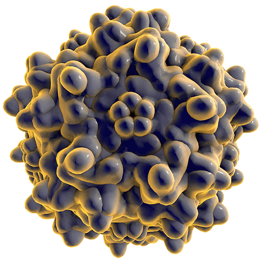
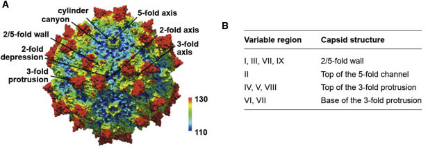 Figure 1. AAV Capsids Share Some Common Structural Features
 Researchers identified Adeno-Associated Virus (AAV), a small single-stranded DNA virus, in 1965 while they were working with adenovirus (AD) when it emerged as a contaminant. The viral structure consists of a 22 nm diameter icosahedral capsid that lacks an envelope. While 80% of humans are seropositive for AAV2 no established human diseases are linked to this virus.
The classification of AAVs relies on their distinct capsid amino acid sequences and their specific binding affinities to different cellular receptors. Eleven serotypes labeled AAV1 to AAV11 have been recognized in primates while additional recombinant isolates have been found in different tissues of both human and non-human primate hosts. The capsid amino acid sequence homology values among serotypes vary between 55% and 99%.
AAV2 stands out as the serotype that scientists have studied most thoroughly because it naturally targets skeletal muscle, neurons, vascular smooth muscle cells, and hepatocytes. This serotype allows for extended transgene expression but produces insufficient transgene activity and faces a ~5 kb packaging constraint when used for liver-targeted hemophilia gene therapy.
AAV5 transports β-galactosidase cDNA to airway and alveolar epithelial cells in mice more effectively than AAV2 and functions without interference from soluble heparin.
Exhibits lower immunogenicity than AAV2. The in vitro studies demonstrate that AAV6 pseudotypes exhibit cellular tropism that differs from AAV2 pseudotypes with AAV6 reaching up to 80% transduction efficiency in airway epithelial cells which makes it beneficial for treating lung diseases like cystic fibrosis through gene therapy.
In mouse models AAV8 provides liver transduction efficiency which exceeds AAV2 by about a factor of 50. The protein shows diminished binding to heparan sulfate because its heparan sulfate-binding area contains reduced basic charge which aligns with its non-heparin-binding behavior. Clinical trials have tested it specifically for liver-targeted gene therapies, which included treatments for hemophilia A and familial hypercholesterolemia.
The single-stranded DNA genome of AAV measures roughly 4.7 kb with 145-nucleotide inverted terminal repeats (ITRs) at each end. Since it does not possess its own polymerase gene AAV requires polymerases from host cells to replicate its genome. The two viral genes rep and cap encode non-structural proteins and structural proteins respectively.
The cap gene generates three distinct capsid proteins named VP1 (87 kDa), VP2 (72 kDa), and VP3 (63 kDa) via alternative splicing and translation initiation. These assemble into a 60-subunit near-spherical capsid. Through two promoters and alternative splicing the rep gene produces four regulatory proteins Rep78, Rep68, Rep52 and Rep40 which function in AAV genome replication.
 Figure 2. AAV genome structure.The AAV genome consists of two parts, rep and cap genes; the ITR is located at both ends (Source: Zhao L, et al. 2024)
Our diverse collection of AAV-related offerings serves scientific research needs across vaccine development projects:
Our product offerings include high-quality AAV antigen products which precisely identify intact viral capsids and customized antigens designed for AAV subunits and replication enzymes. Researchers use these products to identify AAV infections and test vaccine-induced immunity because they deliver high purity combined with strong immunogenicity.
Our AAV antibodies maintain high sensitivity and specificity which makes them perfect for identifying AAV infections and evaluating vaccine effectiveness. These antibodies allow for exact detection of particular AAV antigenic epitopes which makes them excellent research tools.
Our inventory features multiple ELISA kits designed to identify AAV antigenic markers and measure antibody levels. Researchers can achieve precise quantification in AAV studies because these kits deliver accurate results through easy operation.
The titration ELISA kits we provide enable researchers to accurately determine AAV viral titers. These kits enable quick and precise measurement of AAV levels which provides vital information to assist in viral production and experimental setup.
Humans possess immune memory against wild-type AAV. When exposed to AAV capsids the immune system produces antibodies that target them. A range of 1% to 10% of people generate neutralizing antibodies that prevent successful gene transfer. Anti-AAV2 neutralizing antibodies exhibit the highest detection rates between 30% to 60% while showing extensive cross-reactivity across AAV serotypes.
AAV vectors lack viral coding sequences as their antigenic properties come from remaining purification contaminants together with viral capsids and transgene products. The activation of NF-κB and IRF transcription factors results in elevated levels of pro-inflammatory cytokines and type I interferons. During adaptive immune responses antigen-presenting cells (APCs) activate T and B cells by presenting antigens. Effector cells develop from lymphocytes to perform the primary task of eliminating antigens. The presentation of capsid epitopes by transduced cells and APCs through MHC class I molecules to cytotoxic CD8+ T cells results in the removal of AAV-transduced cells while causing inflammation in tissues and diminishing both the transgene expression time frame and its effectiveness.
The construction of recombinant AAV (rAAV) vectors for efficient gene delivery into target cells defines AAV packaging. AAV as a Dependoparvovirus genus member needs a helper virus such as adenovirus or herpesvirus to enable productive infection. The AAV rep and cap genes and the gene of interest along with necessary regulatory elements assemble into viral particles during packaging.
Plasmid Construction
Design plasmids that contain the gene of interest along with ITRs and rep/cap genes. A plasmid that contains adenovirus-derived genes functions as a helper to facilitate AAV replication and packaging.
Cell Transfection
Load the plasmids into a compatible cell line like HEK293 cells through co-transfection. After transfection helper proteins enable AAV genome replication and particle assembly which produces infectious rAAV particles.
Virus Harvesting and Purification
Following an appropriate incubation time cells lysates should be collected and the virus needs to be purified by ultracentrifugation, affinity chromatography or ion-exchange chromatography to achieve high-purity rAAV particles.
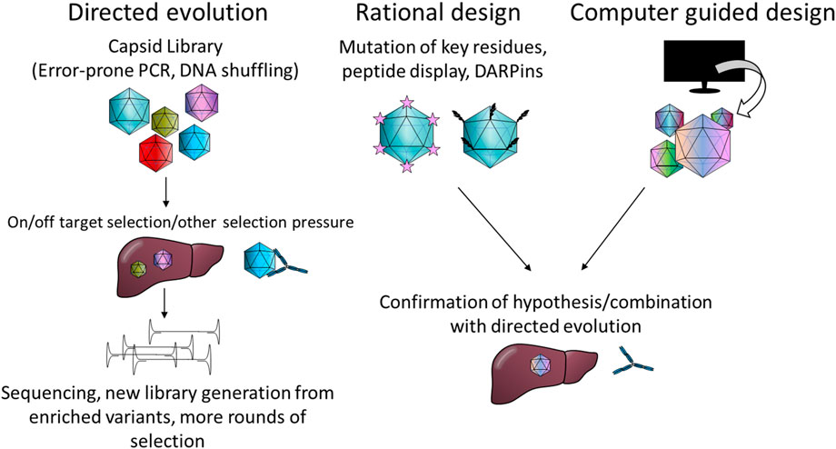 Figure 3. Summary of the most common capsid engineering strategies. (Source: Suoranta T, et al. 2022)
High Efficiency: This process enables target genes to be packaged into viral particles which facilitates effective delivery.
Safety: rAAV does not contain essential replication and integration genes so it stays primarily in the episomal form which reduces potential risks.
Tissue Specificity: The choice of different AAV serotypes enables precise targeting of specific tissues or organs for gene therapy applications.
The gene therapy method using AAV vectors introduces working genes into patient cells to mend or replace their defective genetic material. AAV vectors have the ability to infect both dividing and non-dividing cells while remaining episomal throughout their lifecycle and avoiding integration into the host genome. The transgene produces functional proteins inside the cell which leads to disease correction.
Vector Design and Construction
Select both the therapeutic gene and the suitable AAV serotype for use in vector design. The gene needs placement between AAV ITRs during the rAAV vector construction to achieve strong expression.
Virus Packaging and Production
To maintain high quality and yield outcomes, package the constructed vector into functional AAV particles under strict production conditions.
Patient Administration
Administer the rAAV particles using appropriate delivery methods such as intravenous routes.
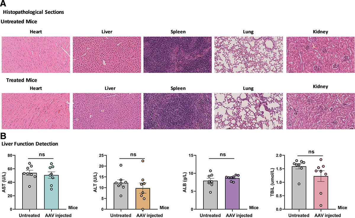 Figure 4. Safety assessment following AAV gene therapy in hemophilia A mice. (A) Histological evaluation (B) Liver function markers (Source: Zhao J-J, et al. 2025)
High Safety: The AAV virus presents no pathogenic threat to humans while provoking only minimal immune system activation. Recombinant AAV does not contain the necessary genes for viral integration and replication which prevents it from integrating into the host genome thereby lowering potential safety risks.
Low Immunogenicity: The low immunogenic nature of AAV vectors enables their persistence within the body without activating significant immune responses thus allowing for sustained gene expression over time.
Tissue-Specific Targeting: Different AAV serotypes enable researchers to target specific tissues or organs thereby improving gene therapy accuracy.
Long-Term Gene Expression: The ability of AAV vectors to remain active in non-dividing cells for extended durations enables extended gene expression periods which makes them effective for treating diseases requiring continuous therapy.
Loading ......
