
Inquiry Basket
 Loading ......
White Spot Syndrome Virus (WSSV) is a rod-shaped virus that infects crustaceans such as shrimp, and belongs to Class II animal diseases. The virus particles are elliptical in shape, with a double layered envelope and a rod-shaped nucleocapsid core. They can be carried by shrimp larvae, contaminated by water sources, and transmitted by planktonic animals. The hosts include various crustaceans such as crabs and lobsters. High temperature, extreme pH values, or chemical treatment can inactivate it. The virus was first discovered in the early 1990s, and research has found that wild crustaceans commonly carry the virus, which can threaten aquaculture populations through vertical transmission. Prevention and control are based on early detection technologies such as PCR, combined with water quality management and shrimp quarantine. The characteristic of virus transmission through aquaculture waste requires strict control over emissions.
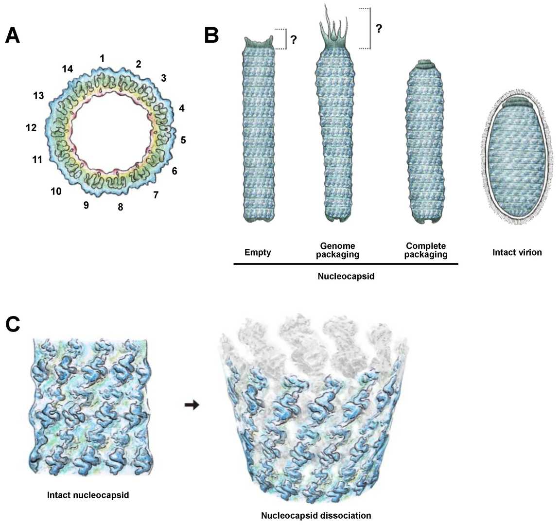 Figure 1. Schematic illustrations of (A) overall C14 symmetry of WSSV nucleocapsid. (Huang, Hui-Ju,et al, 2023)
The pathogenesis of WSSV involves a series of complex interactions between viral proteins and host cell components, controlling virus entry, replication, immune escape, and host cell death. The virus employs a wide range of host manipulation strategies. A series of viral structural proteins, including VP28, play a crucial role in inducing infection and regulating the host environment. These proteins interact with specific host factors to promote the lifecycle of the virus. The following table summarizes the key viral proteins and their interactions in the pathogenesis of WSSV:
| Viral Factor | Category | Key Host Target / Pathway | Function in Pathogenesis |
| VP28 | Major Envelope Protein | Host cell receptor (e.g., Rab7 GTPase), Clathrin-mediated endocytosis pathway. | Serves as the primary attachment and fusion protein. It binds to host receptors to initiate viral entry via endocytosis, making it the foremost target for neutralizing antibodies and a critical diagnostic antigen. |
| WSSV449 (IE1) | Immediate-Early Protein / Transcription Factor | Host RNA polymerase II machinery. | Acts as a master viral transcription factor. It hijacks the host's transcriptional apparatus in the nucleus to drive the expression of viral early genes, effectively launching the viral replication program. |
| Anti-apoptotic Proteins (e.g., WSSV390) | Apoptosis Inhibitors | Host caspase cascade, mitochondrial apoptosis pathway. | Actively inhibit programmed cell death (apoptosis), a key host defense mechanism. By blocking apoptosis, the virus prolongs the survival of infected cells, allowing for greater production of viral progeny. |
Clinical symptoms: Infected shrimp exhibit drowsiness, loss of appetite, and body discoloration. The characteristic signs are white calcifications (0.5-2.0 millimeters) appearing on the inner surfaces of the shell, head, and tail.
Diagnosis: Speculative diagnosis is based on clinical symptoms. The confirmation methods include:
Histopathology: Observation of enlarged nuclei with alkaline inclusions in the ectoderm and mesoderm tissues.
Molecular diagnosis: PCR and nested PCR are the most sensitive and standard methods for early detection.
Immunoassay: Antibody based detection using recombinant VP28 protein as a capture antigen (ELISA, lateral flow test strips) provides a rapid and field deployable diagnostic option.
The virus particle is elliptical in shape and has a capsule with an average size of 430nm x 120nm. The core of the virus particle is an electron dense nucleocapsid, which is rod-shaped with a diameter of 50 urn and an average size of 300 urn x 85 urn. The two ends of the nucleocapsid have cap like structures, with one end being a flattened trapezoid and the other end being a triangular cone. A long tail extends from this end, and the tail has a swollen part. There are 14 spirals between the cap structures, which are perpendicular to the long axis of the nucleocapsid. The vitiligo virus can maintain infectivity in pure water for 1 hour, but loses infectivity in a high salt solution of 3 mol/L NaCl for 1 hour.
White Spot Syndrome Virus is closely related to the microbial community in the intestinal tract of shrimp. When the microbial community is stable and there are many beneficial microorganisms, it can maintain the immune level of shrimp and inhibit virus infection. On the contrary, when the microbial community is unstable and disrupted, it is highly likely to cause white spot disease infection in shrimp. White spot disease caused by deterioration or high water quality indicators (ammonia nitrogen, sub salt, etc.). Weather changes, sudden changes in feed, poor feed quality, and ciliate disease can all cause outbreaks of vitiligo.
White spot syndrome virus is a destructive pathogen, and its economic impact stems from its rapid replication and complex host disruption strategies. The molecular pathogenesis involves synergistic attack: VP28 protein enters through targeted host endocytosis pathway, while viral transcription factors such as IE1 hijack the host nuclear mechanism. Meanwhile, the virus actively inhibits host defense through anti apoptotic proteins and viral miRNA. Recombinant VP28 protein has become indispensable in diagnosis and the cornerstone of vaccine research. Effective management still heavily relies on impeccable biosecurity, and future hope depends on developing effective vaccines and cultivating drug-resistant shrimp populations to address this widespread threat to aquaculture.
Reference
| Target | Cat. No. | Product Name | Host | Isotype | Application | |
| WSSV VP664 | DPATB-H83564 | Anti-WSSV VP664 Polyclonal antibody | Rabbit | IgG | ELISA | Inquiry |
| Target | Cat. No. | Product Name | Host | |
| WSSV VP28 | DAGC571 | Recombinant WSSV VP28 Protein [His] | E. coli | Inquiry |
| WSSV VP19 | DAG-WT782 | Recombinant WSSV VP19 Protein [His] | E. coli | Inquiry |
Loading ......
