Rickettsia is a Gram-negative bacterium that is a type of prokaryotic organism that is obligately parasitic in eukaryotic cells. It is a type of prokaryotic organism that is between bacteria and viruses and close to bacteria. It has no nucleolus and nuclear membrane. Generally spherical or rod-shaped, they are mainly parasitic on arthropods. Some of them can be introduced into the human body through fleas, lice, ticks, and mites, causing diseases such as typhus and trench fever.
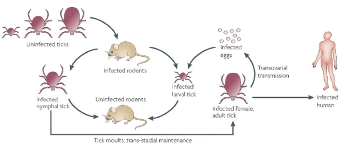 Figure 1. The life cycle of tick-borne rickettsiae. (Walker DH, et al.; 2008)
Rickettsia was first discovered and reported by the young doctor Howard Taylor ricketts in 1906. However, he was unfortunately infected and died while studying Rickettsia. To commemorate his dedication in research and the discovery of Rickettsia For his outstanding contributions, this type of microorganism is named after him.
Rickettsia bacteria are pleomorphic, club-shaped or rod-shaped, with a cell size of 0.3-0.6 μm × 0.8-2.0 μm. Gram staining is negative, but it is not suitable for staining. The outermost layer of the rickettsial body is a mucus layer composed of polysaccharides. Between the mucus layer and the cell wall is a microcapsule composed of polysaccharides and lipopolysaccharides, and then inward are the cell wall and cell membrane. The above-mentioned surface structure is related to bacterial resistance to phagocytosis. There are ribosomes (composed of two subunits, 30S and 50S) in the cytoplasm, and double-stranded DNA in the nucleoplasm, but there is no nuclear membrane and nucleolus. Rickettsia survives in living cells and reproduces by binary fission. It takes 6 to 10 hours to reproduce for one generation. Rickettsia culture is commonly done through animal inoculation, chicken embryo inoculation and cell culture. Commonly used cultured cells include chicken embryo fibroblasts, vero monolayer cells, etc. The optimal culture temperature is 37°C.
Rickettsia has two antigens, namely group-specific antigen and species-specific antigen. The former is related to the lipopolysaccharide component of the cell wall and is heat-resistant. The latter is related to outer membrane proteins and is heat-labile. Rickettsiae have weak resistance and can survive in a few minutes at 56°C. 0.5% phenol and 75% ethanol can kill it in a few minutes. It dies quickly after leaving the host, but can be stored at -20°C or frozen for about half a year, and can survive in the arthropod feces of its medium for more than a year. It is sensitive to tetracycline and chloramphenicol, but sulfa drugs can promote its growth.
Mode of infection: Rickettsia mainly infects the human body through the bites of arthropods such as human lice, rat fleas, and ticks.
Pathogenic substances: Rickettsia are Gram-negative bacteria, and their pathogenic substances are mainly endotoxin and phosphatase. Phosphatase can dissolve the surface mucus layer and microcapsule structure to facilitate adsorption to the host cell surface and resist phagocytosis.
Pathogenic mechanism: After rickettsia invades the body, it first proliferates in local small blood vessel endothelial cells, leading to local inflammatory reaction. The multiplied bacteria enter the bloodstream again to form the first bacteremia, then enter the vascular endothelium in other parts of the body to multiply, and are released into the blood again to form the second bacteremia, resulting in typical clinical symptoms.
Immunity: Rickettsia is an intracellular parasitic bacterium, so the body's immune response is mainly based on cellular immunity. After recovery from infection, patients can develop long-lasting specific immunity to it.
The culture and staining of rickettsiae are important methods for its diagnosis and classification. The most classic method is chicken embryo inoculation, but cell culture can also be used. Staining methods include Giemsa staining, direct immunofluorescence (DFA), etc., but it is difficult to observe positive results under non-culture conditions. The cost of rickettsial pathogenic examination is high and requires high testing personnel and conditions, so it is not used as the main method of clinical testing.
Waifei test: Using the bacterial antigens of Proteus x19, x2 and other strains to replace the rickettsial antigen, and performing a cross-agglutination test with the patient's serum, it can be used to detect whether the patient still has corresponding antibodies. However, this test has been gradually eliminated due to poor specificity, but it is still a classic method for detecting rickettsiae.
Immunofluorescence method: Use immunofluorescence probes to detect the presence of corresponding antibodies in the patient's body to diagnose rickettsial infection. Among them, indirect fluorescence assay (IF) is currently the gold standard for diagnosing rickettsial infection.
Enzyme-linked immunosorbent assay (ELISA): This method has the advantages of being sensitive, simple, and economical. The principle is to use a highly specific immune reaction combined with a highly sensitive enzymatic reaction to detect antigens or antibodies. The antigens coated on its surface are usually recombinant antigens, which have high sensitivity, specificity and reproducibility, and can distinguish between IgG and IgM antibodies.
Polymerase chain reaction (PCR): Fluorescent quantitative PCR can be used to detect rickettsiae at the DNA level. It solves the problem of early detection of rickettsiae and has been adopted by many research institutions.
References
| Target | Cat. No. | Product Name | Expression System | Tag/Conjugate | Application | |
| R. rickettsii Omp | DAG-WT756 | R. rickettsii Omp | Baculovirus | His | N/A | Inquiry |
| DAG-WT757 | R. rickettsii Omp | E. coli | His | N/A | Inquiry | |
| R. typhi | DAGF-008 | R. typhi | E. coli | His | Inquiry | |
| Rickettsia typhi | DAGF-002 | Rickettsia typhi | E. coli | His | WB, ELISA | Inquiry |