


Inquiry Basket
 Loading ......
Threonine proteases are a family of proteolytic enzymes. This family was only recently discovered. Proteases have four major classes based on catalytic mechanism: serine proteases, cysteine proteases, aspartic proteases and metalloproteases. Threonine proteases use an N-terminal threonine residue of its own polypeptide chain as the catalytic nucleophile that cleaves the peptide bond. Threonine proteases account for only about 5% of all proteases but are involved in a variety of important cellular processes. The most representative and functionally important one is the proteasome, which is thought to be the core machinery for protein degradation in eukaryotes, archaea and even some bacteria. The research on proteasomes has also greatly promoted the discovery and research on threonine proteases.
In fact, all of these enzymes are part of a superfamily: N-terminal nucleophile (Ntn) hydrolases. All have a common fold, and a common catalytic mechanism, in which an active N-terminal nucleophilic residue is generated through self-catalytic cleavage.
Different family members employ distinct N-terminal nucleophiles:
Proteasome is the most canonical and well-characterized member of threonine proteases. It is a large multi-subunit barrel-shaped proteolytic complex, which accounts for most of the intracellular protein degradation. The proteins that are primarily degraded by the proteasome are the ones that are targeted by the ubiquitin-proteasome system.
20S Core Particle
This is the proteolytic core of the proteasome. The structure of 20S proteasome is highly conserved and is a stacked assembly of four heptameric rings (α-β-β-α) which forms a hollow barrel-shaped structure with the catalytic active sites present on the inner two β-rings. Following self-cleavage, the individual β-subunits expose their N-terminal threonine residues, which act as the catalytic nucleophiles. The 20S proteasome has multiple peptidase activities, including chymotrypsin-like, trypsin-like and caspase-like activities. It can degrade a broad range of protein substrates.
26S Proteasome
In eukaryotes, the 20S core particle is commonly found with one or two 19S regulatory particles (PA700) bound to its ends. The resulting complex is called the 26S proteasome. The 19S regulatory particle recognizes the polyubiquitin-tagged substrate proteins, and in an ATP-dependent manner unfolds the substrates and translocates them into the catalytic chamber of the 20S core particle for degradation.
Multiple lines of evidence confirm that the proteasome uses the hydroxyl group of its N-terminal threonine as the catalytic nucleophile to cleave peptide bonds:
Mutation Studies
Replacing the catalytic threonine with alanine in archaeal and yeast proteasomes completely abolishes proteolytic activity.
Chemical Modification
The threonine hydroxyl group is modified by irreversible inhibitors (e.g., lactacystin, 3,4-dichloroisocoumarin, vinyl sulfones).
Crystallographic Evidence
X-ray diffraction shows the threonine hydroxyl group forming a hemiactal bond with a peptide aldehyde inhibitor.

Schnell DCABH-229
It was amazing!
Huang DCABH-3342
It's a good product for WB detection
El Bushra DMAB9405
Works well in our assay
Qin DCABH-13708
A very good antibody
Activation of the enzyme and nucleophile: The majority of threonine proteases are produced as proenzymes, that is, in an inactive form. The enzyme is activated when one or several rounds of autocatalytic cleavage (autoproteolysis) are performed, thus unmasking the otherwise deeply buried threonine residue, which is left as the new N-terminus of the polypeptide chain, thus the active site.
Activation of the catalytic nucleophile: Note that in contrast to the classical "catalytic triad" (Ser-His-Asp) present in other serine proteases, the positively charged α-amino group of the enzyme's own N-terminal threonine residue in this instance serves as a general base, i.e. as a proton acceptor, to deprotonate the hydroxyl group (-OH) on the side chain of the adjacent threonine, making it a much better nucleophile.
Nucleophilic attack and acyl-enzyme intermediate formation: The now activated threonine hydroxyl group attacks the carbonyl carbon of the substrate's peptide bond, leading to the unstable formation of the tetrahedral intermediate, after which the peptide bond breaks, releasing the C-terminal peptide fragment and at the same time forming the covalently linked acyl-enzyme intermediate.
Hydrolysis and regeneration of the enzyme: A water molecule enters the active site and is activated (here the α-amino group acts as a general acid by donating a proton) to perform a nucleophilic attack of the carbonyl carbon of the acyl-enzyme intermediate, resulting in the breakage of the covalent bond and release of the N-terminal peptide product. The active site of the enzyme is regenerated and the N-terminal threonine returns to its original state, ready to participate in a new catalytic cycle.
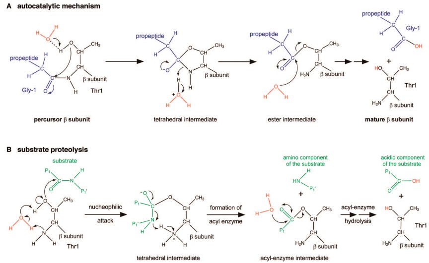 Figure 1. Autocatalytic and proteolytic mechanisms of the 20S proteasome. (Source: Marques AJ, et al. 2009)
By degrading key regulatory proteins, the proteasome precisely controls numerous vital biological processes:
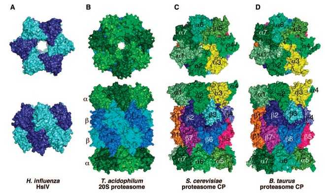 Figure 2. Structure and complexity of pro- and eukaryotic proteasomes. (Source: Marques AJ, et al. 2009)
References
Loading ......
