
Inquiry Basket
 Loading ......
The Herpesviridae are a family of large, double-stranded DNA, enveloped viruses that cause a range of diseases. The nine human herpesviruses are herpes simplex virus 1 (HSV-1), HSV-2, varicella zoster virus (VZV), human cytomegalovirus (HCMV), human herpesvirus 6A (HHV6A), HHV6B, Epstein–Barr virus (EBV), HHV7 and Kaposi sarcoma herpesvirus (KSHV). Herpesvirus infections are ubiquitous and establish lifelong latency in infected hosts. Despite the viruses infecting a variety of cell types, entry into host cells occurs through a conserved mechanism. An understanding of the entry mechanisms for these viruses may provide a basis for the design of antiviral drug candidates and/or subunit vaccines.
Herpesvirus entry into cells requires the coordinated interaction of multiple glycoproteins on the surface of the virion. The initial attachment of a virus to a host cell tethers the virus to the cell, but does not trigger entry. This attachment is mediated by multiple viral glycoproteins and a variety of binding receptors. For herpesviruses, entry receptor binding and membrane fusion functions are performed by multifunctional viral glycoproteins. In the current model of entry, binding to an entry receptor triggers conformational changes in the viral glycoproteins that signal to gB, the fusion protein, to execute membrane fusion. For HSV-1, gD serves the receptor-binding function. gD binding to the receptor prompts an interaction between gD and gH–gL. EBV enters cells in a similar manner, except that gp42 instead of gD serves as the receptor-binding protein for EBV entry into B cells. For HCMV, receptor binding is mediated by two distinct complexes: a trimeric complex including gO and gH–gL or a pentameric complex including UL128, UL130, UL131A and gH–gL. As for HSV-1 and EBV, binding of the HCMV trimer or pentamer to the receptor transmits a signal to gB to trigger fusion.
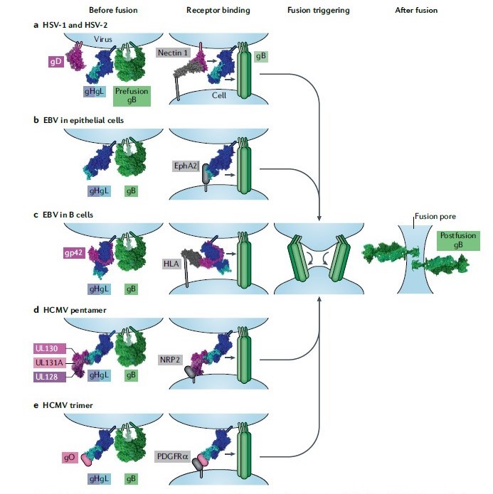 Fig 1. Model of the herpesvirus entry mechanism.
(Nature Reviews Microbiology, 2020)
Herpes simplex viruses 1 and 2 (HSV-1 and HSV-2) fuse with a host cell at the plasma or endosomal membrane. The glycoprotein D (gD) dimer (pink), gH–gL heterodimer (dark and light blue) and gB trimer (green) are necessary and sufficient for entry (column 1). gD binds to one of several entry receptors, including nectin 1 (grey; column 2). Receptor binding displaces the C terminus of the gD ectodomain and transmits a signal to gH–gL (small arrow). gH–gL activates the fusion protein gB (small arrow) to insert hydrophobic fusion loops into the cell membrane. Epstein–Barr virus (EBV) fuses with the plasma membrane of an epithelial cell. gH–gL (blue) and gB (green) are sufficient for fusion (column 1). The binding of gH–gL to ephrin type A receptor 2 (EphA2; grey) triggers gB to insert itself into the host cell (column 2). EBV fusion with B cells occurs in the endosome. A complex of glycoprotein 42 (gp42; pink) and gH–gL (blue) binds to human leukocyte antigen (HLA) class II (grey). The binding triggers gB and may impact membrane orientation (column 2). Human cytomegalovirus (HCMV) entry into epithelial and endothelial cells occurs after endocytosis and requires a pentamer complex of gH–gL (blue) bound to UL128–UL130–UL131A (shades of pink) (column 1). Pentamer binding to neuropilin 2 (NRP2) triggers gB (column 2). HCMV entry into all cells requires a trimer complex comprising gO and gH–gL (column 1). In fibroblasts, the trimer binds to platelet-derived growth factor receptor-α (PDGFRα) and triggers gB at the plasma membrane (column 2). The reason that the trimer is required for entry into epithelial and endothelial cells is currently unclear. After inserting itself into the target cell membrane, gB folds back on itself (column 3). Fusion most likely requires more than one gB trimer to be triggered. The other entry glycoproteins are not included in the figure for clarity. As gB refolds into its postfusion conformation, the viral and cell membranes are fused, creating a fusion pore, through which the viral capsid can enter the cell (column 4).
Reference
Loading ......
