
Inquiry Basket
 Loading ......
Alphavirus is a new important mosquito-borne pathogen from the viral classification Group Ⅳ(+) ssRNA virus family Togaviridae. They spread all over the world and can cause local outbreaks and human epidemics. Alphaviruses can be divided into two independent subgroups: old World (OW) and new world (NW). The OW alphaviruses are loose combinations of polyarthritic viruses endemic to Asia, Europe, Australia, and Africa that contain chikungunya virus (CHIKV), Ross River virus (RRV), Sindbis virus (SINV), and Semliki Forest virus (SFV), which cause human disease. NW viruses usually cause severe encephalitis, including eastern equine encephalitis virus (EEEV), western equine encephalitis virus (WEEV) and Venezuelan equine encephalitis virus (VEEV).
NW alphaviruses cause varying degrees of morbidity and mortality in humans in addition to infecting horses and are important human pathogens. There is a lack of vaccines and antiviral drugs against NW alphaviruses, and the development of therapeutic options could be facilitated by a more detailed molecular understanding of viral replication and host production of infectious viral particles.
Table 1. NW alphaviruses: Geographic distribution, human disease, and route of transmission
| Virus | Endemic Regions | Disease in Humans | Transmission |
| VEEV | United States, Central and South America | Febrile illness, encephalitis; Mortality ~1%; Neurological sequelae in up to 14% of survivors | Mosquito; Aerosol route via laboratory accidents |
| EEEV | North, Central and South America, Caribbean | Febrile illness, encephalitis; Mortality 50%-78%; Neurological sequelae in up to 75% of survivors | Mosquito; Aerosol route via laboratory accidents |
| WEEV | North and South America; Has not been detected in mosquitoes since 2008; | Febrile illness, encephalitis; Mortality 3%-7%; Neurological sequelae in up to 90% of survivors | Mosquito; Aerosol route via laboratory accidents |
(Source: Carey BD, et al. 2019)
The alphavirus genome is divided into two reading frames. The first reading frame encodes the nonstructural polyprotein (nsP), which is further processed into four nonstructural proteins, nsP1-nsP4. The nonstructural proteins form a replication complex that first replicates the viral RNA in a negative-sense, and then transitions it back to the positive-sense RNA, which is incorporated into virion. The second reading frame is controlled by the 26S promoter on the negative-stranded RNA and encodes structural polyproteins. The structural polyprotein consists of the capsid protein and the envelope glycoproteins E1 and E2. nsP3, capsid and E2 proteins have attracted much attention in recent years.
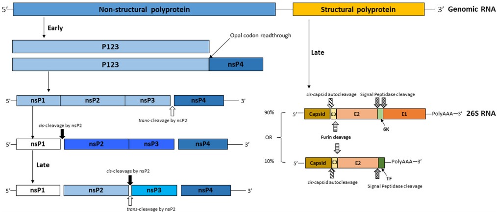 Figure 1. Alphavirus genome and protein coding
(Source: Carey BD, et al. 2019)
Different species of alphaviruses bind to host protein receptors via fusion or viral E2 protein-mediated receptor binding and enter the host cell plasma membrane. Subsequent internalization is mediated by clathrin-mediated endocytosis. In this endocytosis, intracellular vesicles become more acidic during maturation, and the low pH environment leads to dissociation of the E1/E2 dimer, resulting in the insertion of E1 into the endosomal membrane and subsequent fusion of the viral and cellular membranes, releasing the viral nucleocapsid into the cytoplasm of the host. Genomic RNA (gRNA) is immediately translated into two nonstructural precursors, P123 or P1234. p1234 is produced by read-through of the opal termination codon (UGA) between nsP3 and nsP4, followed by cis or trans cleavage of P123 and nsP4, and cis cleavage of nsP1 and P23 only. P123-nsP4 and nsP1-P23-nsP4 utilize host proteins to synthesize negative-stranded viral RNA intermediates through the formation of early-replication complexes. All four mature nonstructural proteins are finally produced between P23, and these late replication complexes are responsible for the synthesis of positive-sense genomic (49S) and subgenomic (26S) RNAs.
The second open reading frame occupies about 1/3 of the alphavirus genome and is controlled by a subgenomic promoter on the negative strand of the viral genome. This ORF encodes the capsid proteins, E3, E2, 6K, and E1 proteins, which are primarily structural proteins that are translated into polyprotein precursors, where the capsid proteins cleave themselves by protein hydrolysis and subsequently encapsidating the gRNAs into the nucleocapsid. This process exposes an ER localization signal on pE2 (the precursor of E3 and E2) that directs the remaining multimeric proteins to the ER, where the proteins enter the secretory pathway for export to the plasma membrane. Later in this pathway, furin-mediated splitting of pE2 forms E2 and E3 proteins, and trimers of E1 and E2 proteins are transported to the plasma membrane.
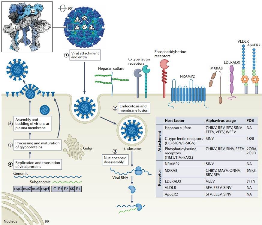 Figure 2. Alphavirus Life Cycle
(Source: Kim AS, et al. 2023)
Although the exact function and mechanism of nsP3's role in viral RNA replication remains to be elucidated, it is generally accepted that it facilitates the assembly of the viral replication complex (vRC) within the host cell. nsP3 is present in the RNA replicase complex and is essential for the synthesis of negative-sense RNAs and subgenomic RNAs. Mutations in the nsP3 coding region adversely affect viral replication, confirming its role in RNA synthesis. nsP3 has three structural domains: the N-terminal macrodomain, the alphavirus unique domain (AUD), and the hypervariable (HVD) C-terminal domain. nsP3's N-terminal macrodomain is considered to be a conserved structure in viruses and is characterized as similar to the human adenosine diphosphate (ADP)-ribose 1′-phosphatase homologue. The AUD domain contains a zinc coordination site consisting of four cysteine residues, where amino acid site mutations make each cysteine residue critical for early RNA replication. The mechanism of HVD action is tentatively unknown, and this region is important for each viral nsP3 to interact with host proteins due to the large sequence differences between alphaviruses. Phosphorylation of HVD in VEEV was found to be ineffective for replication within vertebrate host cells but indispensable for replication within mosquito cells.
Some of the pathways by which nsP3 interacts with host proteins have been revealed, including cytoskeletal reorganization, vesicular transport, stress particle and viral replication formation, RNA binding, and protein synthesis.
The capsid proteins are divided into two separate structural domains, the N- terminal and the C-terminal. The C-terminal is highly conserved in all alphavirus capsid proteins, but the N-terminal domain is highly variable. The capsid proteins have multiple functions in the viral life cycle. The C-terminal domain of the capsid has protease activity and is responsible for releasing the capsid from the translated polypeptide and exposing the ER-localized sequences on the remaining polypeptide so that the glycoprotein can be further processed in the ER. The N-terminal domain is highly positively charged and is responsible for binding viral RNA during assembly. Capsid proteins also play an important role in cellular pathogenicity. The capsid proteins of VEEV and EEEV can lead to host transcriptional turn-off, resulting in cytopathic effects. Further studies have shown that host transcriptional turn-off in VEEV-infected cells is due to the presence of nuclear localization signals (NLS) and nuclear export sequences (NES).
The role of VEEV capsid proteins in host transcriptional turn-off is an important mechanism for evading the innate immune response. In the absence of RanGTP, NES in the N-terminal domain of the capsid interacts with chromosome maintenance 1 (CRM1) (also known as exportin 1). The presence of NLS allows the capsid to bind to the host proteins importin α (Impα) and importin β (Impβ), thereby facilitating the entry of the capsid into the nucleus.
Table 2. Known host protein interactions and inhibitors for capsid
| General Function | Host Protein | Inhibitor | Virus | Cell Type |
| Nuclear Import | Importin α/β | Mifepristone, Ivermectin, G281-1564, 1111684 | VEEV | Vero, MEF |
| Nuclear Export | CRM1 | Leptomycin B, SINE compounds | VEEV, EEEV, WEEV | Vero, MEF |
| Phosphatases | PP1α | 1E7-03 | VEEV, EEEV, WEEV | Vero |
| Kinase, Signal Transduction | PKCδ | Rottlerin | VEEV, EEEV | Vero |
| Ubiquitin proteasome system | undermined | Bortezomib | VEEV, EEEV, WEEV | U87MG, Vero |
(Source: Carey BD, et al. 2019)
The envelope glycoproteins E1 and E2 are processed in the ER after auto-cleavage of the capsid in order to recognize the ER localization sequence in E3. E2 is translated into a polyprotein called pE2, which includes both E3 and E2. Handling pE2 is a critical step in the infection process, as unprocessed pE2 has been shown to affect virus entry. The interaction of E1 and E2 is extremely important for viral budding and cell entry. Both proteins exist as heterodimers in the lipid envelope. E2 is glycosylated and is responsible for receptor-mediated endocytosis. Entry into the cell is via spike E2 components interacting with cell surface receptors. E2 is not only involved in cell attachment, but is also necessary for viral outgrowth, where the assembled nucleocapsid migrates to the cell surface and interacts with the C-terminal cytoplasmic domain of E2. This interaction itself provides sufficient energy for the virus to bud from the cell. Mutations in the E2 gene have also been implicated in epidemiologic virulence determinants and neurotropism of the disease.
Many host components are associated with E2 during viral infection. The actin remodeling pathway is an important component for transporting E2 to the plasma membrane, where E2 binds to actin during infection and co-localizes with the Ras-associated C3 botulinum toxin substrate 1 (Rac1) phosphatidylinositol-4-phosphate 5-kinase type 1 (PIP5K1-α).
References
| Target | Cat. No. | Product Name | Expression System | Tag/Conjugate | Application | |
| VEEV | DAG-WT1030 | Recombinant VEEV E2 glycoprotein (aa 348-701) | E. coli | Unconjugated | Immunogen | Inquiry |
| DAGA-268 | VEEV E2 glycoprotein (aa 348-701) [His] | E. coli | His | WB, ELISA | Inquiry |
| Target | Cat. No. | Product Name | Host | Isotype | Application | |
| WEEV E1 | CABT-CS917 | Mouse Anti-WEEV E1 Monoclonal antibody, clone 3B3D4 | Mouse | IgG2a | IF, FC | Inquiry |
| CABT-CS916 | Mouse Anti-WEEV E1 Monoclonal antibody, clone 3B4E6 | Mouse | IgG2a | IF, FC | Inquiry | |
| CABT-CS915 | Mouse Anti-WEEV E1 Monoclonal antibody, clone 3B7D8 | Mouse | IgG2b | IF, FC | Inquiry | |
| WEEV E2 | CABT-B1090 | Anti-WEEV E2 polyclonal antibody | Rabbit | IgG | ELISA | Inquiry |
| Target | Cat. No. | Product Name | Expression System | Tag/Conjugate | Application | |
| WEEV | DAGA-269 | WEEV E2 glycoprotein (aa 1-366) [His] | E. coli | His | WB, ELISA | Inquiry |
| WEEV E2 | DAGB177 | Recombinant Western Equine Encephalitis Virus E2 GP [His] | Mammalian cells | His | WB | Inquiry |
| DAGB176 | Western Equine Encephalitis Virus E2 GP [His] | Insect cells | His | WB | Inquiry |
| Target | Cat. No. | Product Name | Host | Isotype | Application | |
| EEEV | CABT-CS919 | Humanized Anti-EEEV Monoclonal antibody, clone 249 | Humanized | IgG1 | ELISA, Neut | Inquiry |
| EEEV E1 | CABT-CS914 | Mouse Anti-EEEV E1 Monoclonal antibody, clone 2C2D5 | Mouse | IgG2b | ELISA, IF, HI | Inquiry |
| CABT-CS913 | Mouse Anti-EEEV E1 Monoclonal antibody, clone 2B5C7 | Mouse | IgG2b | ELISA, IF, HI | Inquiry | |
| EEEV E2 | CABT-CS918 | Humanized Anti-EEEV E2 Monoclonal antibody, clone 230 | Humanized | IgG1 | ELISA, Neut | Inquiry |
| CABT-B1088 | Anti-EEEV E2 Glycoprotein polyclonal antibody | Rabbit | IgG | ELISA, WB | Inquiry |
| Target | Cat. No. | Product Name | Expression System | Tag/Conjugate | Application | |
| EEEV | DAGA-267 | EEEV E2 glycoprotein (aa 338-687) [His] | E. coli | His | WB, ELISA | Inquiry |
| DAGB175 | Eastern Equine Encephalitis Virus E3E2 Protein [His] | Insect cells | His | WB | Inquiry |
Loading ......
