
Inquiry Basket
 Loading ......
Viruses in the family Polyomaviridae are found in a wide range of host species, since the first polyomavirus was isolated and identified as mouse K virus in 1952, 73 viruses have been identified, of which 13 have been found in humans. JC polyomavirus (JCPyV), the first human polyomavirus isolated from patients with the initials JC in 1971, is highly prevalent in people around the world, with a serum positive rate of between 40% and 60%. It is the only human polyomavirus known to cause neurological diseases.
JCPyV is a non-enveloped icosahedral virus with a diameter of 40nm, which can cause hemagglutination (HA) of human type 0 erythrocytes, which provides a means for seroepidemiological research. JCVPyV has been found around the world that a large proportion of people have seroconversion before adulthood, and healthy people, including pregnant women, produce anti-JCV immunoglobulin G (IgG). Its genome is a closed circular and superhelical double-stranded DNA, and its capsid is composed of three viral structural proteins: VP1, VP2 and VP3, of which VP1 is the main component. The virus has 72 pentamers, each of which consists of five VP1 molecules and one VP2 or VP3 molecule. Only VP1 is exposed to the surface of the capsid, so it determines the specificity of the receptor. Polyomavirus DNA is a nucleosome structure, with about 25 nucleosomes composed of virus DNA and host cell histone. Virions do not contain linker histones and are obtained when they enter the host cell.
The prototype of JCPyV genome has 5130bp, but the length of each variant is different due to the change of non-coding region. The genome encodes six major viral proteins (large T antigen and small T antigen, VP1, VP2, VP3 and agnoprotein) and several splicing variants of T antigen. The early and late transcripts of the genome are physically separated by non-coding control regions (NCCR) and transcribed in the opposite direction from the opposite DNA strands. The early part of the genome transcribed before the beginning of DNA replication consists of large T antigen and small T antigen genes and splicing variants T'135, T'136 and T'165. The transcription of the late genome is carried out simultaneously with DNA replication, and encodes three kinds of viral structural proteins (VP1, VP2 and VP3) as well as accessory agnoprotein. Large T antigen and its variants can interact not only with host protein and virus protein, but also with host DNA and virus DNA. T protein drives host cells into the S phase of virus replication, regulates the transcription of host and virus genomes, and directly participates in viral DNA replication.
 Figure 1. Schematic diagram of the JCV genome
(Source: Ferenczy MW, et al. 2012)
JCPyV is transmitted by the oral route and excreted in urine and feces. In people with normal immune function, primary JCPyV infection has no obvious symptoms, and then the virus lurks in certain tissues, such as bone marrow, brain, tonsils, kidney cells and lungs. The researchers call this dormant JCPyV the archetype plant, from which other JCPyV variants evolved. Typical JCPyV cannot replicate effectively in macroglia, but rearranges genes in the non-coding regulatory region of its genome and transforms into neurotropic prototype JCPyV associated with oligodendrocyte infection and central nervous system (CNS) diseases. Its genome contains rearrangements, including repetition, tandem repetition, insertion, and deletion, and is the most commonly found form of JCPyV in cerebrospinal fluid (CSF) and brain tissue of patients with PML. In the case of persistent immunosuppression, the prototype variation of JCPyV will lead to productive infection in the central nervous system. It is not clear whether the prototype variant originated from the brain or peripheral tissue, or whether it originated from both the brain and peripheral tissue. How the immune system prevents the virus from replicating and spreading to CNS, and how immune damage allows the virus to spread in the white matter of CNS, have not been accurately explained.
JCPyV does not cause significant symptoms in normal individuals, whereas it causes CNS disease in immunosuppressed or immunodeficient individuals. Prior to the advent of highly effective antiretroviral therapy for AIDS, the most common immunosuppressed cause of JCPyV-associated CNS disease lesions was AIDS, and the most common manifestation was progressive multifocal leukoencephalopathy (PML). PML is a lethal neurodegenerative disease characterized by lysogenic infection of oligodendrocytes and astrocytes in the CNS by JCPyV. Following cell lysis, JCPyV spreads to neighboring regions causing focal destruction of oligodendrocytes, leading to demyelination, and the lack of myelin leads to axonal dysfunction, which may ultimately result in the permanent loss of neurons. PML lesions in the brain affect white matter and tend to be multifocal, suggesting that the virus spreads to the brain via the hematogenous route. Symptoms of PML are characterized by motor dysfunction, visual deficits, and language deficits, but a clinical diagnosis requires adjunctive tests such as magnetic resonance imaging (MRI) of the brain to visualize characteristic multifocal lesions and the presence of JCPyV DNA in the CSF or the presence of JCPyV proteins and DNA in a brain biopsy in order to complete an accurate diagnosis.
At present, it is believed that the hypothesis of PML is that latent JCPyV must occur in at least four ways to cause lytic infection of oligodendrocytes in the brain: (I) the host immune system must be impaired or changed, and (ii) virus NCCR must obtain changes that increase viral transcription and replication in B cells and glial cells, (iii) DNA binding factors that bind to recombinant NCCR sequence motifs must be present and / or up-regulated in infected hematopoietic progenitor cells, B cells and / or glial cells, (iv) viruses in free viruses or B cells must cross the blood-brain barrier and be brought into the brain, where the virus is transmitted to oligodendrocytes and lytic infection occurs.
 Figure 2. JCPyV genome undergoes rearrangement in PML
(Source: Atkinson AL, et al. 2020)
JCPyV initially binds to target cells via α2,6-linked glycan lactoseries tetrasaccharide c (LSTc), a receptor motif that has both the requisite α2,6-linked silicic acid and a distinctive L shape. At this early stage, JCPyV may also interact with adipocyte plasma membrane associated protein (APMAP), a surface protein found in glial cells and throughout the body. JCPyV also interacts transiently with the 5- hydroxytryptamine 2 receptor (5-HT2R), which is then clathrin-mediated endocytosed into the cell and subsequently translocated to Rab-5-positive early endosomes. After entering the cell, JCPyV is transported to the endoplasmic reticulum (ER), and the viral capsid begins to degrade under the action of the resident protein of ER. Then the virus uses ER-related degradation pathway to shuttle from ER to the cytoplasm and finally into the nucleus, where the internal DNA is released in the nucleus and begins to replicate.
One problem in the above description of virus entry pathway is that glial cells in the brain do not express LSTc receptors and do not bind to viruses, and JCPyV cannot rely on LSTc receptors to enter cells. One hypothesis is that the virus can invade cells using a non-receptor infection mode, in which case the virus does not need to maintain the function of the receptor and does not worry about the spread of mutations in the receptor site. Extracellular vehicles (EVs) can contain anything including proteins, nucleic acids, and lipids to transmit information between different cells. Several viruses, including HIV-1, HSV-1 and HepA viruses, have used EV to transmit virus particles between cells. JCPyV is one of the viruses that utilize EVs, both externally bound and packaged internally, and these virus-associated EVs are highly infectious to cultured glial cells in a receptor-independent manner that is also protected from neutralizing antibodies.
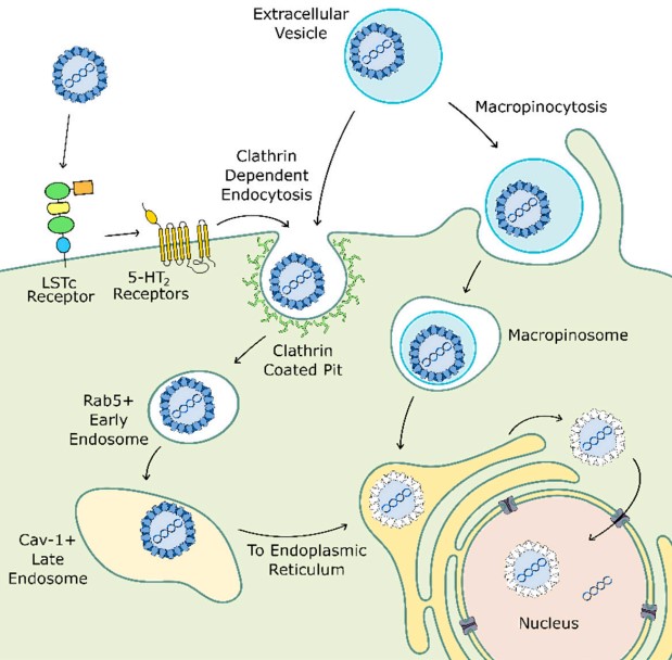 Figure 3. JCPyV cellular entry may occur through multiple pathways
(Source: Atkinson AL, et al. 2020)
After reaching the nucleus, JCPyV expresses its early genes to stop the cell in S phase, and recruits the cell DNA damage response protein to the local region adjacent to the cell promyelocytic nuclear bodies (called the virus assembly factory). With the transcription of late JCPyV genes, VP1 tubules begin to form in the nucleus and JCPyV agnoprotein is produced, which facilitates DNA replication and promotes virus release through a mechanism similar to that of other virus pore proteins.
References
| Target | Cat. No. | Product Name | Size | Species | Application | Detection Sample | |
| JCV VP1 | DEIASL160 | Anti-polyomavirus JC (JCV) IgG ELISA Kit | 96T | Human | Quantitative | Serum and plasma | Inquiry |
| Target | Cat. No. | Product Name | Host | Isotype | Application | |
| JCV VP1 | DMABT-Z59741 | Anti-JCV VP1 Monoclonal antibody | Mouse | IgG2a | I-ELISA, WB | Inquiry |
| Polyomavirus early antigen | DMABT-Z60972 | Anti-Polyomavirus Early Antigen Monoclonal antibody, Clone RaE | Rat | IgG2b | WB, IP, ICC, IF | Inquiry |
| Polyomavirus Large T antigen | DMABT-Z59919 | Anti-Polyomavirus Large T antigen Monoclonal antibody, Clone RaLV | Rat | IgG2b | WB, IP, ICC, IF | Inquiry |
| Polyomavirus Medium T | DMABT-Z60955 | Anti-Polyomavirus Medium T Monoclonal antibody, Clone RaOV | Rat | IgG2b | IP, ICC, IF, WB | Inquiry |
| Polyomavirus PyMT | DMAB7134 | Anti-Polyomavirus PyMT Monoclonal antibody, Clone Hmv-Hmv | Mouse | IgG1 | IF, WB, IP | Inquiry |
| Target | Cat. No. | Product Name | Expression System | Tag/Conjugate | Application | |
| JCV VP1 | DAG-P2868 | JCV Major Capsid VP1 (full length) | S. cerevisiae | Unconjugated | ELISA, WB, SDS-PAGE | Inquiry |
| Polyomavirus Major Capsid VP1 | DAG-P2867 | Polyomavirus Major Capsid VP1 (full length) | S. cerevisiae | Unconjugated | ELISA, WB, SDS-PAGE | Inquiry |
| DAG-P2869 | Polyomavirus Major Capsid VP1 (full length) | S. cerevisiae | Unconjugated | ELISA, WB, SDS-PAGE | Inquiry |
Loading ......
