
Inquiry Basket
 Loading ......
Mucin is a macromolecular glycoprotein, including two major categories: secretory and membrane-bound. It is mainly synthesized by epithelial cells, and a small part exists on hematopoietic cells. A large number of studies have shown that mucin is crucial to the occurrence and development of tumors. Mucin 1 (MUC1) is a membrane-bound mucin, which is abnormally expressed and glycosylated in malignant tumors of important organs such as the gastrointestinal tract, liver, pancreas, lung and breast. It participates in cell biological activities such as signal transduction and immune regulation, and plays a key role in tumor infiltration, metastasis, proliferation, apoptosis and inflammation.
MUC1 is expressed on the apical membrane of epithelial cells and is encoded by the 1q21 gene. It contains two subunits: MUC1 N-terminus (MUC1-N) and MUC1 transmembrane C-terminus (MUC1-C). MUC1-N includes a variable number of tandem repeats (VNTR) and sea urchin sperm protein enterokinase and agrin domains. VNTR is composed of an amino acid peptide sequence (PDTRPAPGSTAPPAHGVTSA) rich in proline, threonine and serine residues (PTS domain). MUC1-C is composed of an extracellular domain of 58 amino acids, a transmembrane domain of 28 amino acids and a cytoplasmic domain of 72 amino acids, which can anchor MUC1-N to the cell surface. Under normal conditions, MUC1 undergoes autoproteolytic cleavage in the SEA module within the tandem repeat sequence, thereby producing MUC1-N and MUC1-C2 subunits, which form a heterodimer complex through stable hydrogen bonds and exist on the plasma membrane; under the catalysis of shedding enzymes, the complex dissociates, MUC1-C and MUC1-N are released, and it also catalyzes the cleavage of the extracellular structure of MUC1-C. The dissociated MUC1-C enters the nucleus of tumor cells and plays a role in promoting the growth, proliferation, invasion and migration of tumor cells and other malignant behaviors.
Glycosylation is one of the important post-translational modifications of proteins, and changes in glycosylation are necessary for tumor growth and progression. There are abundant O-glycosylation sites on serine and threonine residues in the variable number tandem repeat sequence region (VNTR sequence) of MUC1-N, while the degenerate sequence of MUC1-N contains N-glycosylation sites, which are O-glycosylated and N-glycosylated in the Golgi apparatus and endoplasmic reticulum, respectively. O-glycosylation is related to the biological properties of MUC1, while N-glycosylation plays an important role in protein folding, secretion and apical expression.
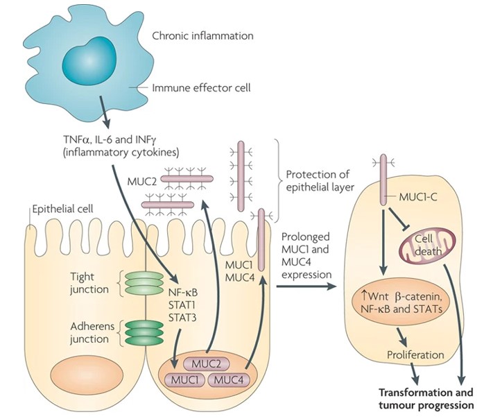 Figure 1. Mucins, chronic inflammation and cancer. (Sources: Kufe DW., 2009)
In the fully glycosylated state, the abundant sugar chains can cover the ligand binding sites and isolate signal factors to prevent them from activating receptors. On tumor cells, branched glycans are lost, ligand binding sites are exposed, and various protein-protein interactions activate signal pathways; or specific branched sugar chains (certain protein binding sites) are lost, which hinders signal transduction.
MUC1 in normal cells undergoes high glycosylation, so the immunogenic epitopes in the VNTR region are covered by extensively branched sugar chains, protecting them from the extracellular environment. In tumor cells, MUC1 protein is often abnormally hypoglycosylated, allowing these immunogenic epitopes to be exposed to the immune system. Currently, these epitopes have become potential targets for tumor immunotherapy. Tumor-associated mucin glycosylation also plays a role in immune escape. When Tn and STn antigens are formed, they bind to macrophage galactose-specific lectins expressed by macrophages, drive immunosuppressive programs, induce effector T cell apoptosis, and promote tumor immune escape. Glycosylation of MUC1 induces the differentiation of immune cells and the production of factors related to tumor progression. The ST antigen formed by sialylation of O-linked sugars on MUC1 has the potential to bind to Siglec-9 in the sialic acid-binding immunoglobulin-like lectin (Siglecs) family expressed on monocytes and macrophages. After binding to monocytes, ST antigens induce a pro-inflammatory phenotype, which can recruit immune cells into tumors, induce monocytes to differentiate into macrophages and dendritic cells, and induce the secretion of factors associated with tumor progression in a Siglec-9-dependent manner.
Anoikis is a special form of apoptosis that maintains tissue homeostasis by removing displaced epithelial or endothelial cells and preventing them from being seeded in inappropriate locations. In tumor cells, MUC1 is overexpressed, and O-glycosylation modification on its extracellular structure prevents the interaction of cell surface anoikis-initiating molecules (such as integrins, E-cadherin, and Fas) with ligands, resulting in resistance to anoikis. Some scholars have conducted experimental studies and found that inhibiting MUC1 O-glycosylation by inhibiting the expression of core-1 glycosyltransferase (C1GT) can increase the anoikis of MUC1-positive tumor cells.
In a large number of cancer and precancerous lesion studies, it was found that the abundant sugar chains on MUC1 are often modified by sialic acid, galactose, fucose, N-acetylglucosamine and N-acetylgalactosamine, forming complex branched sugar chains that can serve as attachment sites for pathogenic microorganisms and as a potential source of nutrition. Salmonella invades intestinal epithelial cells through the mucus layer and causes infection. MUC1, as a component of the intestinal apical transmembrane mucin and mucus layer, promotes the invasion of Salmonella. Salmonella adhesin SiiE protein was identified as participating in MUC1-mediated apical invasion of gastrointestinal epithelial cells. It contains a series of tandem repeat sequences that interact with the tandem repeat sequences of MUC1 in a zipper-like manner. This SiiE-MUC1 interaction is required for the destruction of the mucin barrier and promotes the entry of Salmonella into intestinal epithelial cells. The O-linked glycans on the tandem repeats of MUC1 undergo sialic acid modification, which can become the main target of Salmonella macroadhesin SiiE and promote the interaction between MUC1 and SiiE protein.
The cytoplasmic tail of MUC1-C has multiple phosphorylation sites, which interact with various intracellular signaling proteins to activate or mediate cell signal transduction, and participate in tumor infiltration, metastasis, proliferation and apoptosis.
EGFR is a member of the epidermal growth factor receptor tyrosine kinase (ErbB) family, including EGFR/ErbB1 (Her1), ErbB2 (Her2/c-Neu), ErbB3 (Her3) and ErbB4 (Her4), which has the function of regulating cell growth and differentiation. When the proto-oncogene mutates, EGFR is overexpressed, causing uncontrolled cell proliferation. MUC1 is expressed on the apical membrane of epithelial cells, while EGFR is expressed on the basolateral membrane of epithelial cells. In malignant tumor cells and when epithelial stress occurs, cell polarity is lost, and MUC1-C and EGFR are relocated and expressed throughout the cell membrane to form a complex, promoting the activation of downstream signal transduction and the upregulation of MUC1-C expression; when the extracellular domain of MUC1-C is phosphorylated, it can serve as a binding site for galectin-3. In the presence of EGFR ligand, the binding of galectin-3 to the extracellular domain of MUC1 can induce polarization of the MUC1 cell surface and increase the MUC1-EGFR interaction, leading to increased homo/heterodimerization and activation of EGFR. In addition, MUC1 can increase EGFR promoter activity and EGFR mRNA and protein levels, inhibit EGFR degradation and mediate EGFR localization in the nucleus, affecting the interaction between EGFR and the transcriptionally active promoter region. Currently, treatment of breast cancer cells with EGFR inhibitors can lead to upregulation of circulating MUC1 protein expression in plasma, so MUC1 has been used as a sensor to monitor the effect of EGFR targeted therapy on tumors.
PI3Ks belong to the lipid kinase family. MUC1 can regulate the expression and signal transduction of various tyrosine kinase receptors that transmit signals through the phosphoinositide 3-kinase (PI3K) pathway, including EGFR and STAT3, and plays an important role in driving cell proliferation, migration and survival. The cytoplasmic tail of MUC1-C contains a YHPM sequence, which can serve as a direct binding site for PI3K after phosphorylation. It interacts with the Src homology 2 (SH2) domain of the PI3Kp85 subunit, which can change the conformation of p85, thereby activating the PI3Kp110 catalytic subunit and then activating the PI3KAKT pathway; the cell-penetrating peptide GO-203 can block the interaction between MUC1-C and PI3Kp85 and inhibit the phosphorylation of the complex composed of Akt and its downstream effector mTOR. Matrix metalloproteinase 2/9 (MMP-2/9) is an important downstream target of the PI3K-Akt signaling pathway in tumors and is involved in tumor cell migration and invasion. Studies have found that silencing the MUC1 gene reduces the protein levels of PI3K, Akt and downstream targets MMP-2/9 in tumor cells, blocking or inhibiting tumor invasion and metastasis.
β-catenin is a downstream molecule of the Wnt signaling cascade and usually forms a complex with cell surface adhesion molecules such as E-cadherin, leading to stable cell-cell interactions between epithelial cells. Under normal circumstances, intracellular β-catenin is phosphorylated by glycogen synthase kinase 3b (GSK3b). Phosphorylated MUC1-C directly binds to β-catenin, stabilizes the β-catenin/TCF4 complex, and promotes the activation of Wnt target genes such as cyclin D1 and MYC, thereby affecting the transcription and carcinogenesis of these target genes. In addition, MUC1 enhances the activation of β-catenin and p120catenin and affects the subcellular localization of p120catenin. The three can jointly regulate the conduction of the WNT signaling pathway. Studies have shown that the classical Wnt/b-catenin pathway is one of the pathways involved in the mesenchymal epithelial transition (EMT) process. MUC1 activates β-catenin and binds to the promoter WRE of SNAIL (a transcription factor associated with EMT), resulting in an increase in the nuclear translocation of the SNAIL transcription factor, thereby regulating EMT.
The NF-κB pathway is a common inflammatory pathway. Activation of this pathway can induce the expression of inflammatory cytokines. After MUC1-C is introduced into the nucleus, the cytoplasmic tail directly binds to NF-κBp65 through the GGSSLSY motif to form a complex and increases the proportion of NF-κB on the promoter of the target gene, thereby activating the NF-κB pathway; after the inflammatory pathway is activated, the expression of inflammatory factors such as tumor necrosis factor α (TNF-α) and interleukin 6 (IL-6) in inflammatory and tumor cells is upregulated. At the same time, TNF-α in turn activates the binding of NF-κBp65 to the MUC1κB promoter site, inducing the expression of the MUC1 gene in tumor cells. The positive feedback loop of the two interacts with each other and accelerates tumor development. MUC1-C can also be associated with the NF-κB pathway by promoting the activation of the TAK1 promoter mediated by NF-κB, inducing TAK1 expression and forming a complex with MUC1-C, further promoting the binding of TAK1 and TRAF6.
JNK is a member of the mitogen-activated protein kinase (MAPK) family, which can intervene in oncogene transformation, cell apoptosis and cell proliferation, and participate in the occurrence and development of tumors. JNK includes three types: JNK1, JNK2 and JNK3. MUC1-C can directly bind to JNK1, enhance the activation of JNK1 and c-Jun under genotoxic stress caused by chemotherapeutic drugs such as cisplatin, and block the apoptosis of colon tumor HCT116 cells to DNA damage. In hepatocellular carcinoma, MUC1 activates the JNK/AP-1 pathway, which in turn mediates autocrine transforming growth factor β (TGF-β) signaling, further leading to the phosphorylation of Smad2C. In addition, after JNK activation, it directly phosphorylates Smad2L, and phosphorylated Smad2L and Smad2C enhance MMP-9 expression, mediating the migration and invasion of liver cancer cells.
ER includes two major categories: ER-α and ER-β. ER-α is present in large quantities in breast tissue, while ER-β is mainly expressed in the intestine. MUC1-C can directly bind to the ER-α DNA binding domain. After the interaction between the two, they exist in the ER-α transcription complex, inducing ER-α-mediated gene transcription and promoting estrogen-mediated breast cancer cell growth. In addition, ER-α is also involved in other cell signaling pathways, and its related non-transcriptional mechanisms trigger the PI3K/AKT signaling pathway, and the continuous activation of this signaling pathway blocks cell apoptosis. ER-β has been found to play an anti-inflammatory and anti-tumor role in the colon. The disease activity of colitis caused by ER-β deficiency is significantly increased, and the reduction of the protective mucus layer and the dedifferentiation of related colon tissues are related to the upregulation of MUC1.
References
| Target | Cat. No. | Product Name | Host | Isotype | Application | |
| CA 27-29 | DMAB6139 | Anti-CA 27-29 monoclonal antibody, clone N4021210 | Mouse | IgG1 | ELISA, WB | Inquiry |
| Target | Cat. No. | Product Name | Expression System | Tag/Conjugate | Application | |
| CA 27-29 | DAGA6120 | CA 27-29 protein | Human | Unconjugated | N/A | Inquiry |
Loading ......
